


When it comes to skin growths, understanding the distinction between actinic keratosis and seborrheic keratosis is crucial for proper treatment and health management. While these conditions may sound similar, they have different causes, appearances, and potential health risks that require distinct approaches to care.
Understanding Actinic Keratosis
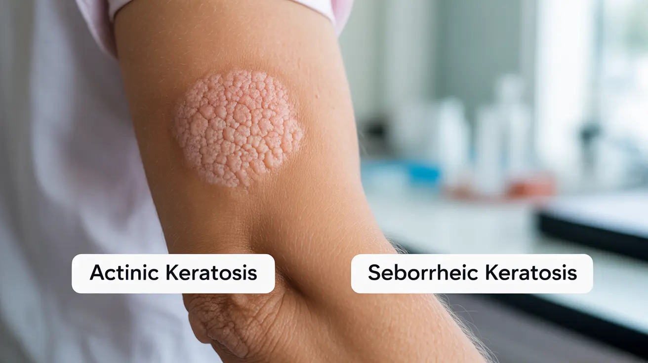
Actinic keratosis (AK) represents a precancerous skin condition that develops due to prolonged sun exposure. These rough, scaly patches typically appear on sun-exposed areas of the body and require medical attention due to their potential to develop into skin cancer.
Characteristics and Symptoms of Actinic Keratosis
Actinic keratosis lesions commonly present as:
- Rough, scaly patches that feel like sandpaper
- Pink, red, or brown coloration
- Size ranging from 2-6 millimeters
- Sometimes accompanied by itching or burning
- Most frequently found on face, ears, neck, and hands
Understanding Seborrheic Keratosis
Seborrheic keratosis (SK) is a benign skin growth that commonly develops with age. Unlike actinic keratosis, these growths are not precancerous and typically don't require medical intervention unless they cause discomfort or cosmetic concerns.
Key Features of Seborrheic Keratosis
Seborrheic keratosis typically appears as:
- Waxy, stuck-on appearance
- Light tan to dark brown color
- Well-defined borders
- Varying sizes from very small to over an inch wide
- Can appear anywhere on the body except palms and soles
Key Differences in Diagnosis and Risk Factors
The primary distinction between these conditions lies in their underlying causes and potential health risks. Actinic keratosis develops from UV damage and requires monitoring due to cancer risk, while seborrheic keratosis is largely genetic and age-related with no cancer risk.
Treatment Approaches
Managing Actinic Keratosis
Treatment for actinic keratosis is essential and may include:
- Cryotherapy (freezing)
- Topical medications
- Photodynamic therapy
- Regular skin checks
- Strict sun protection measures
Addressing Seborrheic Keratosis
Treatment for seborrheic keratosis is usually optional and may involve:
- Cryotherapy for removal
- Curettage or shave excision
- Electrocautery
- Laser therapy for cosmetic purposes
Prevention Strategies
While seborrheic keratosis cannot be prevented, actinic keratosis can be avoided through proper sun protection measures including:
- Daily sunscreen use (SPF 30 or higher)
- Protective clothing and hats
- Avoiding peak sun hours
- Regular skin examinations
Frequently Asked Questions
What is the difference between actinic keratosis and seborrheic keratosis in terms of symptoms and risks?
Actinic keratosis appears as rough, scaly patches that can develop into skin cancer, while seborrheic keratosis presents as waxy, stuck-on growths that are benign. AK is caused by sun damage and carries cancer risk, while SK is genetic and age-related with no cancer risk.
How do you treat actinic keratosis effectively to prevent it from becoming cancerous?
Effective treatment options include cryotherapy, topical medications, photodynamic therapy, and consistent sun protection. Regular monitoring by a healthcare provider is essential to prevent progression to skin cancer.
Can seborrheic keratoses be dangerous or lead to any serious health issues?
Seborrheic keratoses are generally harmless and don't lead to serious health issues. While they may cause irritation or cosmetic concerns, they don't develop into skin cancer.
What are the common causes and risk factors for developing actinic keratosis?
The main risk factors include cumulative sun exposure, fair skin, older age, weakened immune system, and a history of sunburns. People who spend significant time outdoors or use tanning beds are at higher risk.
How can I distinguish between actinic keratosis and seborrheic keratosis based on their appearance and medical diagnosis?
Actinic keratosis appears as rough, scaly patches that feel like sandpaper and are usually pink or red. Seborrheic keratosis has a waxy, stuck-on appearance and is typically brown. A healthcare provider can make a definitive diagnosis through visual examination or biopsy if needed.


