


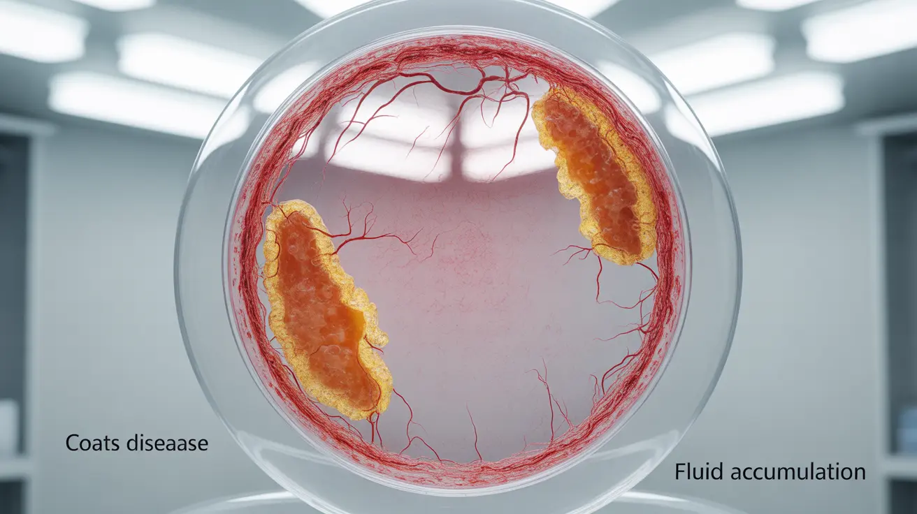
Coats eye disease is a rare but serious retinal condition that primarily affects children and young adults. This disorder occurs when abnormal blood vessels develop in the retina, leading to fluid buildup and potential vision problems. Early detection and proper management are crucial for preserving vision and achieving the best possible outcomes.
While this condition can be concerning for parents and patients, understanding its signs, symptoms, and treatment options can help ensure prompt medical attention and appropriate care. Let's explore the key aspects of Coats eye disease, including how it's diagnosed and managed.
Signs and Symptoms of Coats Eye Disease
Recognizing the early warning signs of Coats eye disease is essential for timely intervention. Common symptoms include:
- Vision loss or decreased vision in one eye
- Crossed eyes (strabismus)
- White glow in the pupil (leukocoria)
- Poor depth perception
- Eye redness or inflammation
These symptoms typically appear gradually and often affect only one eye. Since young children may not be able to articulate vision problems clearly, regular eye examinations are crucial for early detection.
Diagnosis Process
Diagnosing Coats eye disease involves several specialized tests and examinations performed by eye care professionals. The diagnostic process typically includes:
Initial Eye Examination
An ophthalmologist will conduct a thorough eye examination using specialized equipment to view the retina and blood vessels.
Advanced Imaging Tests
- Fluorescein angiography
- Optical coherence tomography (OCT)
- Ultrasound imaging
- Fundus photography
Treatment Approaches
Treatment for Coats eye disease varies depending on the severity and stage of the condition. Available options include:
Laser Therapy
Light laser treatment can help seal leaking blood vessels and reduce fluid accumulation in the retina.
Cryotherapy
This freezing treatment targets abnormal blood vessels to prevent further leakage and damage.
Advanced Interventions
In severe cases, additional treatments may be necessary, such as:
- Intravitreal injections
- Surgery for retinal detachment
- Drainage procedures for excess fluid
Prevention and Management
While Coats eye disease cannot be prevented, proper management can help minimize its impact:
- Regular eye examinations
- Early intervention when symptoms appear
- Consistent follow-up care
- Monitoring for potential complications
Frequently Asked Questions
What are the early signs and symptoms of Coats eye disease to watch for in children?
Early signs include a white glow in the pupil, crossed eyes, vision loss in one eye, and poor depth perception. Parents should be particularly vigilant if they notice their child having difficulty with depth perception or showing signs of vision problems in one eye.
How is Coats eye disease diagnosed and what tests are commonly used?
Diagnosis involves comprehensive eye examinations, fluorescein angiography to examine blood vessel patterns, optical coherence tomography (OCT) to view retinal layers, and ultrasound imaging. These tests help doctors assess the extent of the condition and plan appropriate treatment.
What treatment options are available for Coats eye disease at different stages?
Treatment options include laser photocoagulation for early stages, cryotherapy to freeze abnormal blood vessels, and surgical interventions for advanced cases. The choice of treatment depends on the disease stage and severity of symptoms.
Can Coats eye disease lead to permanent vision loss or blindness if untreated?
Yes, without proper treatment, Coats eye disease can progress to severe vision loss or blindness in the affected eye. Early detection and intervention are crucial for preventing these serious complications.
Is Coats eye disease hereditary or related to other systemic health problems?
Coats eye disease is not typically hereditary and usually occurs sporadically. While it's generally not associated with other systemic health conditions, some rare genetic conditions may have similar presentations, making proper diagnosis important.


