


When diagnosing and monitoring Chronic Obstructive Pulmonary Disease (COPD), chest X-rays serve as valuable diagnostic tools that can reveal characteristic changes in the lungs. Understanding the differences between COPD X-rays and normal chest X-rays can help both healthcare providers and patients better comprehend the progression of this chronic respiratory condition.
In this comprehensive guide, we'll explore the key features that distinguish COPD chest X-rays from normal chest X-rays, and discuss why these differences matter for diagnosis and treatment planning.
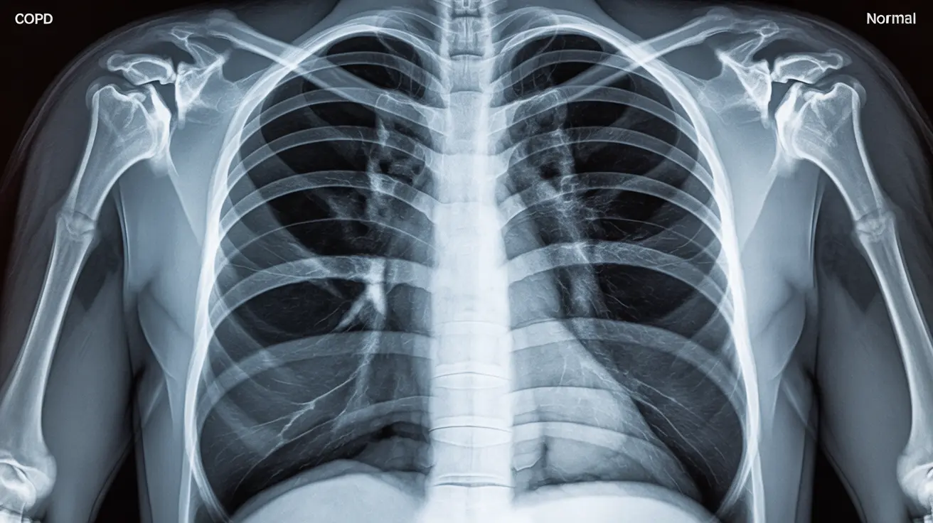
Key Features of COPD on Chest X-rays
COPD creates distinct patterns on chest X-rays that trained medical professionals can identify. These changes reflect the underlying damage to lung tissue and airways that characterizes the disease.
Hyperinflation Patterns
One of the most notable differences in COPD X-rays is the appearance of hyperinflated lungs. This presents as increased darkness and enlarged lung fields on the X-ray image. The lungs appear larger than normal, and the space between ribs often appears wider due to the trapped air within the lungs.
Diaphragm Changes
In COPD patients, the diaphragm typically appears flattened and sits lower in the chest cavity compared to normal X-rays. This flattening occurs due to the increased pressure from hyperinflated lungs, making it harder for the diaphragm to function normally during breathing.
Structural Changes in COPD X-rays
Bronchial Wall Thickening
COPD X-rays often show thickened bronchial walls, appearing as prominent linear markings throughout the lung fields. This thickening results from chronic inflammation and tissue remodeling in the airways.
Bullae Formation
Advanced COPD may reveal bullae - large air spaces caused by the destruction of lung tissue. These appear as dark, round areas on the X-ray and indicate severe emphysema, a common component of COPD.
Limitations of Chest X-rays in COPD Diagnosis
While chest X-rays can show significant COPD changes, they have certain limitations that healthcare providers must consider when making diagnostic decisions. Early-stage COPD may not be visible on standard chest X-rays, which is why additional testing is often necessary.
Role of CT Scans
Computed Tomography (CT) scans provide more detailed images of lung tissue and can detect COPD changes earlier than traditional X-rays. They're particularly useful for identifying subtle emphysema patterns and assessing the distribution of disease throughout the lungs.
Frequently Asked Questions
What are the main differences between a COPD chest X-ray and a normal chest X-ray?
COPD chest X-rays typically show hyperinflated lungs, flattened diaphragms, increased lung transparency, and wider spaces between ribs. Normal chest X-rays display proper lung size, a curved diaphragm, and normal tissue density.How does COPD cause changes like lung hyperinflation and diaphragm flattening on an X-ray?
COPD causes air trapping in the lungs, leading to hyperinflation. This trapped air pushes down on the diaphragm, causing it to flatten. The increased pressure also creates wider spaces between ribs and changes the overall appearance of the chest cavity.Can a normal chest X-ray rule out early stages of COPD?
No, a normal chest X-ray cannot definitively rule out early-stage COPD. Early changes may be too subtle to detect on standard X-rays, which is why pulmonary function tests and other diagnostic tools are essential for accurate diagnosis.Why are CT scans sometimes preferred over X-rays for diagnosing COPD?
CT scans provide more detailed, three-dimensional images of lung tissue, allowing healthcare providers to detect subtle changes and early-stage COPD that might not be visible on standard X-rays. They also better show the distribution and severity of emphysema.What do bullae or air pockets seen on a COPD X-ray indicate about lung damage?
Bullae on X-rays indicate severe emphysema, where lung tissue has been destroyed, creating large air-filled spaces. Their presence suggests advanced COPD and significant damage to the lung's air sacs, affecting breathing capacity.


