


If you're experiencing persistent back pain, your healthcare provider may recommend a CT scan to help identify the underlying cause. CT (computed tomography) scans use advanced X-ray technology to create detailed cross-sectional images of your spine, providing valuable insights for diagnosis and treatment planning.
Understanding what to expect from a CT scan for back pain can help ease any concerns and ensure you're properly prepared for the procedure. This comprehensive guide covers everything from preparation to results interpretation.
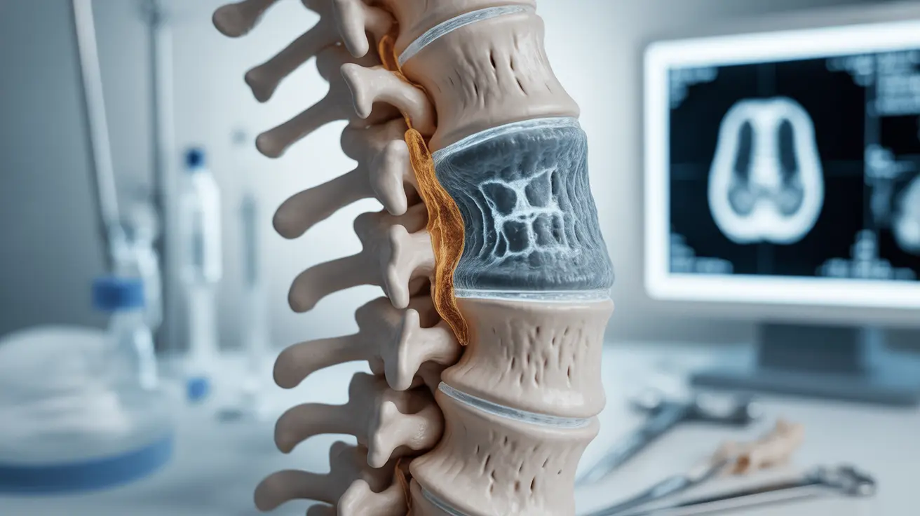
Understanding CT Scans for Back Pain Diagnosis
A CT scan combines multiple X-ray images taken from different angles to create detailed 3D pictures of your spine and surrounding tissues. This advanced imaging technique is particularly effective at visualizing bone structures and can help identify various conditions causing back pain.
Common Conditions Detected by Spinal CT Scans
CT scans are especially useful for diagnosing:
- Herniated discs
- Spinal fractures
- Bone spurs
- Spinal stenosis
- Tumors or abnormal growths
- Vertebral compression
- Signs of infection
Preparing for Your Spinal CT Scan
Proper preparation ensures the best possible imaging results. Your healthcare provider will provide specific instructions, but general preparation typically includes:
- Removing metal objects, including jewelry
- Wearing comfortable, loose-fitting clothing
- Fasting for several hours if contrast material will be used
- Informing your doctor about medications and allergies
- Notifying staff if you're pregnant or might be pregnant
What to Expect During the Procedure
The CT scan process is quick and non-invasive. You'll lie on a table that slides into a donut-shaped machine. The scanner rotates around you, capturing images from multiple angles. The entire procedure usually takes 15-30 minutes, though actual scanning time is much shorter.
CT Scan vs. MRI for Back Pain
While both imaging techniques are valuable diagnostic tools, they serve different purposes. CT scans excel at showing bone detail and are typically faster and more widely available than MRIs. They're particularly useful for detecting fractures and evaluating bone-related conditions.
Safety and Risk Considerations
CT scans are generally safe, but they do involve exposure to ionizing radiation. The benefits of accurate diagnosis typically outweigh the minimal risks. Your healthcare provider will carefully consider whether a CT scan is the most appropriate imaging option for your specific situation.
Frequently Asked Questions
What conditions can a CT scan detect to explain back pain?
A CT scan can detect various conditions including herniated discs, spinal fractures, bone spurs, spinal stenosis, tumors, and infections. It's particularly effective at showing detailed bone structure and identifying structural abnormalities that may be causing pain.
How should I prepare for a lumbar spine CT scan for back pain?
Preparation typically involves removing metal objects, wearing comfortable clothing, and possibly fasting if contrast material will be used. Inform your healthcare provider about any medications, allergies, or potential pregnancy before the procedure.
What are the differences between a CT scan and an MRI for diagnosing back pain?
CT scans are better at showing bone detail and are faster than MRIs. MRIs provide superior soft tissue imaging and don't use radiation. CT scans are often preferred for evaluating bone-related conditions, while MRIs are better for examining soft tissues, nerves, and discs.
Is a CT scan safe for back pain diagnosis and what are the risks?
CT scans are generally safe but do involve exposure to ionizing radiation. The radiation dose is carefully controlled and monitored. The benefits of accurate diagnosis typically outweigh the minimal risks associated with radiation exposure.
How long does a CT scan for lower back pain take and what can I expect during the procedure?
A CT scan typically takes 15-30 minutes total, with actual scanning time being much shorter. You'll lie still on a table that moves through a donut-shaped scanner. The procedure is painless, though you may need to hold specific positions briefly for optimal imaging.


