


An ERG eye test, or electroretinography, is a sophisticated diagnostic procedure that measures the electrical responses of various cell types in the retina, including photoreceptors (rods and cones) and other neural cells. This important test helps eye care professionals evaluate how well your retina functions and can detect various retinal disorders, even before visible symptoms appear.
Whether you're preparing for an upcoming ERG test or seeking to understand this diagnostic tool better, this comprehensive guide will explain everything you need to know about the procedure, its importance, and what to expect.
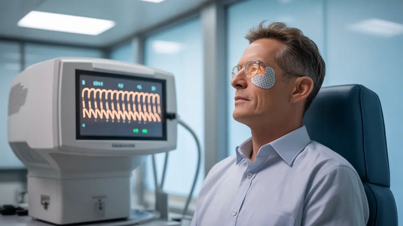
How an ERG Eye Test Works
During an ERG eye test, specialized equipment measures your retina's response to light stimulation. The test uses electrodes, typically in the form of contact lenses or small threads, to record the electrical activity generated by your retina's cells when exposed to different light conditions.
The procedure captures two main types of responses:
- Scotopic responses (testing rod function in dark conditions)
- Photopic responses (testing cone function in light conditions)
Test Components and Equipment
The main equipment used in an ERG eye test includes:
- Special contact lens electrodes or thread-like sensors
- A device that produces specific light patterns
- Recording equipment to measure electrical responses
- A display screen for real-time monitoring
Importance of ERG Testing
ERG eye tests serve as crucial diagnostic tools for various retinal conditions. They can help identify and monitor:
- Inherited retinal disorders
- Age-related retinal diseases
- Toxic retinopathies
- Inflammatory retinal conditions
- Various forms of color blindness
Clinical Applications
Eye care professionals often recommend ERG testing when:
- Evaluating unexplained vision loss
- Screening for inherited retinal diseases
- Monitoring disease progression
- Assessing treatment effectiveness
- Conducting pre-surgical evaluation
Preparing for Your ERG Eye Test
Proper preparation ensures accurate test results. Your doctor may advise:
- Avoiding caffeine for 24 hours before the test
- Removing contact lenses
- Coming with clean skin around the eyes (no makeup)
- Following specific dark adaptation instructions
- Bringing someone to drive you home
The Testing Process
The ERG eye test typically follows these steps:
- Pupil dilation
- Dark adaptation period (usually 20-30 minutes)
- Electrode placement
- Light stimulus exposure
- Data recording and analysis
Post-Test Recovery
After the ERG eye test, you may experience:
- Temporary light sensitivity
- Mild discomfort where electrodes were placed
- Blurred vision from dilating drops
These effects typically resolve within a few hours.
Frequently Asked Questions
What does an ERG eye test measure and how is it performed? An ERG eye test measures the electrical responses of different retinal cells to light stimulation. It's performed by placing specialized electrodes on or near the eye and recording the retina's electrical activity when exposed to various light patterns.
Why is the ERG eye test important for diagnosing retinal diseases? The ERG eye test is crucial because it can detect retinal dysfunction before visible symptoms appear. It provides objective measurements of retinal cell function, helping doctors diagnose and monitor various retinal conditions accurately.
How should I prepare for an electroretinography (ERG) test? Prepare by avoiding caffeine 24 hours before the test, removing contact lenses, arriving without eye makeup, and arranging transportation home. Your doctor may provide additional specific instructions based on your situation.
What should I expect during and after the ERG eye test procedure? During the test, expect pupil dilation, a dark adaptation period, electrode placement, and exposure to various light patterns. Afterward, you may experience temporary light sensitivity and mild discomfort that typically resolves within hours.
What do abnormal ERG test results indicate about retinal health? Abnormal ERG results may indicate various retinal conditions, including inherited disorders, degenerative diseases, or toxic effects on the retina. The specific pattern of abnormality helps doctors determine the type and extent of retinal dysfunction.


