


Pregnancy ultrasounds are essential medical imaging tools that help healthcare providers monitor both maternal and fetal health throughout pregnancy. These non-invasive procedures use sound waves to create detailed images of your developing baby, providing crucial information about growth, development, and overall well-being.
Whether you're preparing for your first ultrasound or wanting to learn more about these important prenatal screenings, understanding what to expect can help ease any anxiety and prepare you for these significant pregnancy milestones.
Types of Pregnancy Ultrasounds
Healthcare providers may recommend different types of ultrasounds throughout your pregnancy, each serving specific purposes:
Transvaginal Ultrasound
Often performed during early pregnancy (before 12 weeks), this internal ultrasound provides detailed images of the developing embryo, confirms pregnancy location, and helps establish accurate dating.
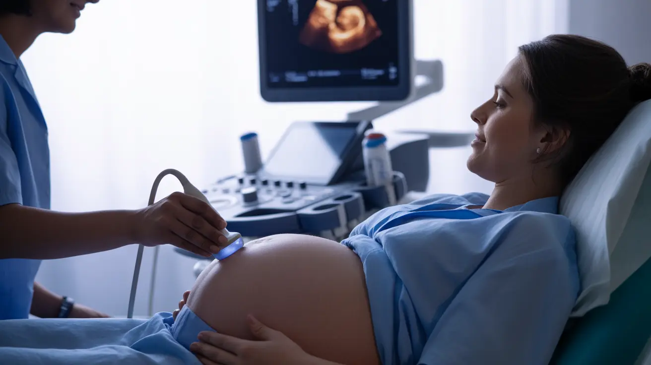
Transabdominal Ultrasound
This common external ultrasound becomes the primary imaging method after the first trimester, offering comprehensive views of fetal development, placental position, and amniotic fluid levels.
3D and 4D Ultrasounds
These specialized scans create detailed three-dimensional images or real-time videos of your baby, though they're typically not medically necessary and are often done for keepsake purposes.
Timing of Pregnancy Ultrasounds
Most pregnant women can expect several ultrasounds throughout their pregnancy:
- First Trimester (6-13 weeks): Dating scan and viability check
- Second Trimester (18-22 weeks): Detailed anatomy scan
- Third Trimester: As needed to monitor growth and position
Safety and Benefits
Pregnancy ultrasounds have been used safely for decades and provide numerous benefits:
- Confirming pregnancy and due date
- Monitoring fetal growth and development
- Detecting potential complications early
- Determining baby's position before delivery
- Checking placental location and health
Preparing for Your Ultrasound
Preparation requirements vary depending on the type and timing of your ultrasound:
- Early pregnancy scans may require a full bladder
- Wear comfortable, two-piece clothing
- Follow any specific instructions from your healthcare provider
- Consider bringing a support person
- Be prepared to lie still for 30-60 minutes
What Ultrasounds Can Reveal
Modern ultrasound technology provides detailed information about:
- Fetal heartbeat and movement
- Number of babies
- Gender (if desired and visible)
- Growth measurements
- Developmental milestones
- Potential health concerns
Frequently Asked Questions
What is the purpose of a pregnancy ultrasound and when is it typically performed during pregnancy?
Pregnancy ultrasounds are performed to monitor fetal development, confirm pregnancy dating, and screen for potential complications. They're typically scheduled during early pregnancy (6-13 weeks), mid-pregnancy (18-22 weeks), and as needed in the third trimester.
How safe are ultrasounds for the pregnant mother and developing baby?
Ultrasounds are considered very safe when performed by trained healthcare professionals. They use sound waves rather than radiation and have been safely used in pregnancy for over 50 years with no known risks to mother or baby.
What are the different types of pregnancy ultrasounds and how do they differ?
The main types include transvaginal ultrasounds (internal, early pregnancy), transabdominal ultrasounds (external, throughout pregnancy), and 3D/4D ultrasounds (specialized imaging). Each type serves different purposes and is used at specific stages of pregnancy.
What can an ultrasound reveal about the baby's health and development?
Ultrasounds can reveal fetal heartbeat, size, position, number of babies, gender, major anatomical structures, and potential health concerns. They also assess placental location, amniotic fluid levels, and overall fetal well-being.
How should I prepare for a pregnancy ultrasound and what happens during the procedure?
Preparation may include having a full bladder for early scans and wearing comfortable clothing. During the procedure, gel is applied to your abdomen (or a probe is inserted vaginally for early scans), and a technician moves the ultrasound device to capture images. The procedure typically takes 30-60 minutes.


