


The heel is far more than just the back part of your foot – it's a complex anatomical structure that serves as the foundation for your entire body's weight-bearing system. Understanding heel anatomy is essential for recognizing how this remarkable area supports movement, absorbs impact, and maintains stability throughout daily activities.
From the largest bone in your foot to the intricate network of tendons, ligaments, and soft tissues, the heel's anatomy is perfectly designed to handle the tremendous forces placed upon it with each step. Whether you're walking, running, or simply standing, your heel works tirelessly to keep you mobile and stable.
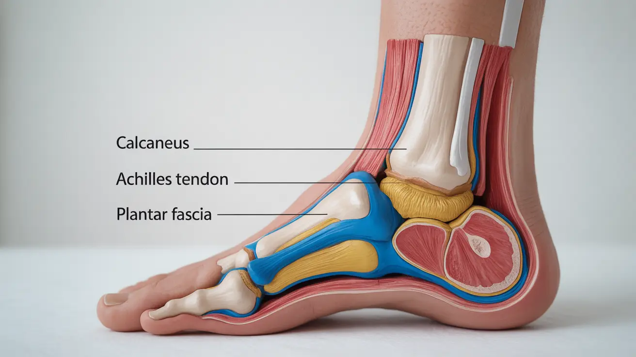
The Calcaneus: Your Heel's Primary Foundation
At the core of heel anatomy lies the calcaneus, also known as the heel bone. This substantial bone serves as the largest bone in the human foot and forms the primary structural foundation of the heel region. The calcaneus is uniquely designed with a robust, rectangular shape that can withstand significant compressive forces.
The calcaneus plays multiple critical roles in foot function. Its broad surface area provides an excellent base of support for the entire body, while its strategic positioning allows it to serve as a lever arm for the powerful muscles of the calf. The bone's internal structure features a honeycomb-like pattern of trabecular bone that helps distribute weight evenly and absorb shock during impact.
Several important anatomical landmarks on the calcaneus contribute to its functionality. The posterior tuberosity serves as the attachment point for the Achilles tendon, while the sustentaculum tali supports the talus bone above. These features work together to create a stable platform for movement and weight distribution.
The Achilles Tendon Connection
The Achilles tendon represents one of the most critical components of heel anatomy, connecting the powerful calf muscles to the calcaneus. This thick, fibrous cord is the strongest tendon in the human body, measuring approximately 6 inches in length and capable of withstanding forces several times body weight.
The connection between the Achilles tendon and heel bone occurs at the posterior tuberosity of the calcaneus. This attachment point is crucial for movement, as it allows the calf muscles to pull on the heel bone during activities like walking, running, and jumping. The mechanical advantage created by this lever system enables efficient propulsion and helps generate the force needed for forward movement.
The Achilles tendon's role extends beyond simple movement generation. It also helps control the foot's position during the stance phase of walking, preventing excessive dorsiflexion and contributing to overall stability. The tendon's elastic properties allow it to store and release energy, improving the efficiency of locomotion.
Shock Absorption and Weight Distribution Systems
The heel anatomy includes several specialized structures designed to absorb shock and distribute body weight effectively during walking and other activities. The heel fat pad, located beneath the calcaneus, serves as the primary shock-absorbing cushion. This specialized tissue contains chambers filled with fat cells that compress under load and spring back to their original shape.
The heel fat pad's unique construction allows it to absorb up to 80% of the impact forces generated during heel strike. This remarkable capability protects not only the heel bone but also the joints and structures throughout the lower extremity and spine. The fat pad's thickness and density can vary among individuals, influencing overall shock absorption capacity.
Additionally, the calcaneus itself contributes to shock absorption through its internal structure. The trabecular bone pattern acts like a natural spring system, compressing slightly under load and then returning to its original configuration. This bone-level shock absorption works in conjunction with the soft tissue structures to create a comprehensive impact management system.
The Plantar Fascia and Heel Connection
The plantar fascia represents a crucial component of heel anatomy, consisting of a thick band of fibrous tissue that extends from the calcaneus to the toes. This structure originates from the medial tubercle of the calcaneus and fans out across the bottom of the foot, creating a strong supportive network.
The plantar fascia's connection to the heel bone is fundamental to maintaining the foot's arch structure. Acting like a bowstring, it helps support the longitudinal arch of the foot and prevents excessive flattening during weight-bearing activities. This support is essential for efficient walking mechanics and proper force distribution across the foot.
During the push-off phase of walking, the plantar fascia experiences significant tension as it helps propel the body forward. The windlass mechanism, created by the interaction between the plantar fascia and the metatarsals, generates additional arch support and contributes to the foot's natural spring-like properties. This mechanism is crucial for energy conservation during locomotion.
Supporting Ligaments and Stabilization
Multiple ligaments within the heel anatomy work together to provide stability and support during standing and walking activities. The spring ligament, also known as the plantar calcaneonavicular ligament, connects the calcaneus to the navicular bone and helps support the medial longitudinal arch.
The long plantar ligament extends from the calcaneus to the cuboid and metatarsal bones, providing support for the lateral aspect of the foot. This ligament works in conjunction with the plantar fascia to maintain arch integrity and distribute forces effectively across the foot's structure.
The calcaneofibular ligament connects the calcaneus to the fibula, contributing to ankle stability and preventing excessive inversion of the foot. Similarly, the deltoid ligament complex on the medial side of the ankle provides stability and helps control foot position during various activities.
These ligamentous structures work as an integrated system, providing both static support when standing and dynamic stability during movement. Their proper function is essential for maintaining normal foot mechanics and preventing injury during daily activities.
Frequently Asked Questions
What is the calcaneus and what role does it play in foot function?
The calcaneus is the largest bone in the foot, commonly known as the heel bone. It serves as the primary foundation for the heel and plays several critical roles in foot function, including providing a stable base for weight-bearing, serving as an attachment point for the Achilles tendon, and acting as a lever arm for calf muscle action. Its robust structure and strategic positioning make it essential for walking, running, and maintaining balance.
How does the Achilles tendon connect to the heel and why is it important for movement?
The Achilles tendon connects to the heel by attaching to the posterior tuberosity of the calcaneus (heel bone). This connection is crucial for movement because it allows the powerful calf muscles to pull on the heel bone, creating the force needed for walking, running, and jumping. The tendon acts as a lever system that enables efficient propulsion and helps control foot position during various activities.
What structures in the heel help absorb shock and distribute body weight when walking?
The primary shock-absorbing structure in the heel is the heel fat pad, which sits beneath the calcaneus and can absorb up to 80% of impact forces during heel strike. Additionally, the internal trabecular bone structure of the calcaneus itself acts like a natural spring system, compressing under load and returning to its original shape. These structures work together to protect the heel bone and distribute forces throughout the foot and lower extremity.
Why is the plantar fascia important and how does it connect to the heel bone?
The plantar fascia is a thick band of fibrous tissue that connects to the heel bone at the medial tubercle of the calcaneus and extends to the toes. It's important because it supports the foot's longitudinal arch, prevents excessive flattening during weight-bearing, and contributes to efficient walking mechanics through the windlass mechanism. This connection helps maintain proper foot structure and assists in forward propulsion during walking and running.
What ligaments support the heel and help stabilize the foot during standing and walking?
Several ligaments support the heel and provide foot stability, including the spring ligament (plantar calcaneonavicular ligament), which connects the calcaneus to the navicular bone and supports the medial arch; the long plantar ligament, which extends from the calcaneus to the cuboid and metatarsals; and the calcaneofibular ligament, which connects the heel to the fibula for ankle stability. These ligaments work together as an integrated system to provide both static support when standing and dynamic stability during movement.


