


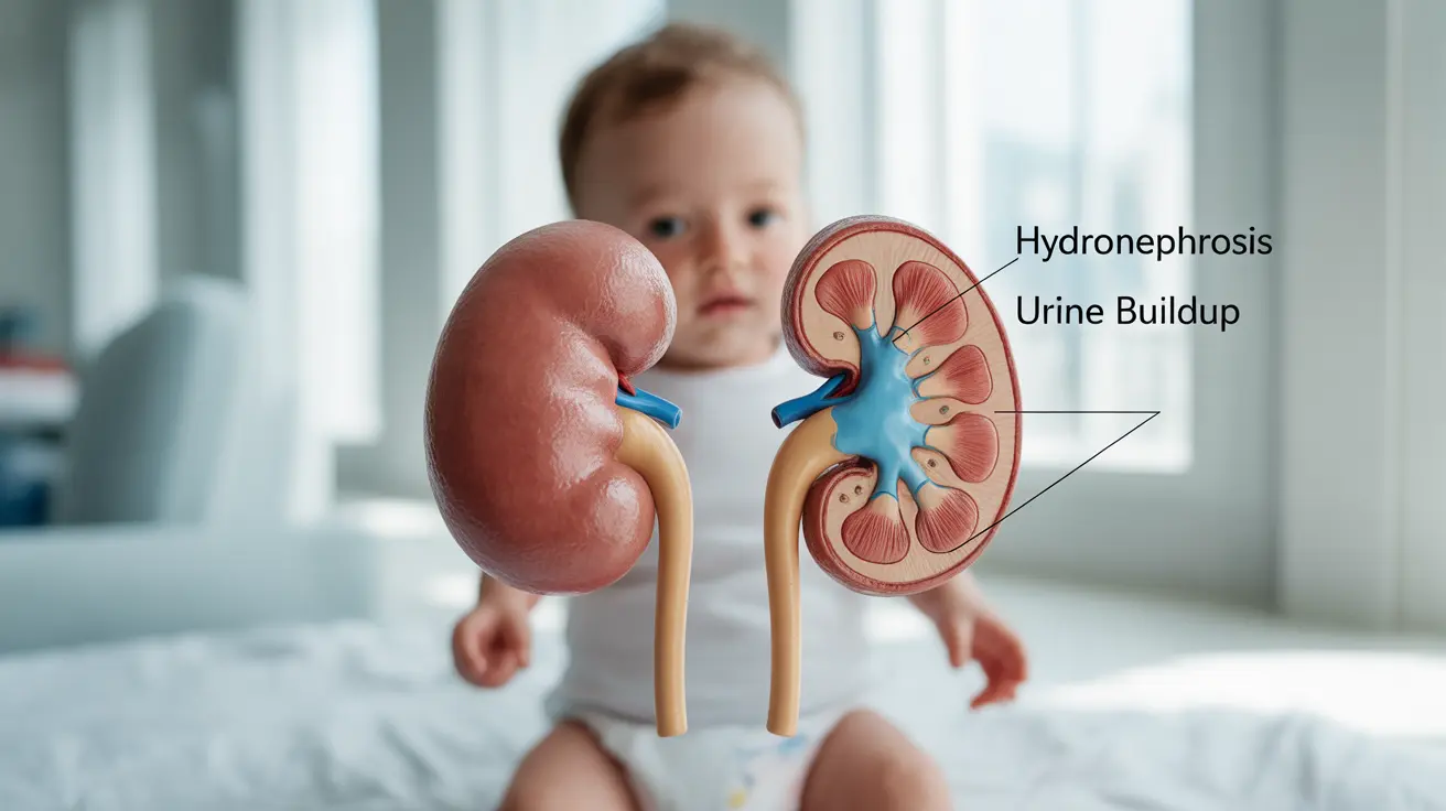
Hydronephrosis in infants is a condition where one or both kidneys become swollen due to a buildup of urine. This common urinary tract condition can be detected during prenatal ultrasounds and affects approximately 1 in every 100 to 200 pregnancies. Understanding this condition is crucial for parents and caregivers to ensure proper medical attention and the best possible outcomes for affected infants.
While the diagnosis of hydronephrosis can be concerning for parents, it's important to note that many cases, especially mild ones, may resolve on their own without intervention. However, proper medical evaluation and monitoring are essential to prevent potential complications and ensure appropriate treatment when necessary.
Prenatal Detection and Causes
Hydronephrosis is often first discovered during routine prenatal ultrasounds, typically during the second or third trimester of pregnancy. The condition can affect one kidney (unilateral) or both kidneys (bilateral), and its severity can vary significantly.
Common Causes
Several factors can lead to hydronephrosis in infants:
- Ureteropelvic junction (UPJ) obstruction
- Vesicoureteral reflux (VUR)
- Posterior urethral valves (in male infants)
- Ureterocele
- Structural abnormalities in the urinary tract
Signs and Symptoms
While some infants with hydronephrosis may not show any obvious symptoms, others might display various signs that warrant medical attention:
- Poor feeding or irritability
- Urinary tract infections
- Abdominal mass
- Poor urine stream
- Crying during urination
- Failure to thrive
Diagnostic Procedures
When hydronephrosis is suspected or confirmed, healthcare providers typically employ several diagnostic tools to assess the condition's severity and determine the best course of action:
Primary Diagnostic Tests
- Ultrasound imaging
- Voiding cystourethrogram (VCUG)
- Nuclear medicine scans
- Blood tests to assess kidney function
- Urinalysis to check for infections
Treatment Approaches
The treatment plan for infant hydronephrosis depends on various factors, including the condition's severity, underlying cause, and the presence of complications. Treatment options may include:
Conservative Management
For mild cases:
- Regular monitoring through ultrasound
- Preventive antibiotics when necessary
- Watchful waiting for spontaneous resolution
Surgical Intervention
For severe cases or when conservative management isn't effective:
- Pyeloplasty
- Valve ablation
- Ureterocele repair
- Ureteral reimplantation
Frequently Asked Questions
What causes hydronephrosis in infants and how is it detected before birth?
Hydronephrosis in infants is typically caused by blockages or abnormalities in the urinary system. It's usually detected during routine prenatal ultrasounds, where technicians can observe enlarged kidneys or dilated collecting systems.
What are the common signs and symptoms of hydronephrosis in newborns and infants?
Common signs include poor feeding, irritability, urinary tract infections, abdominal masses, and abnormal urination patterns. However, some infants may not show any obvious symptoms, especially in mild cases.
How is hydronephrosis in infants diagnosed and what tests are typically used?
Diagnosis involves ultrasound imaging, VCUG, nuclear medicine scans, blood tests, and urinalysis. These tests help determine the severity of the condition and identify any underlying causes.
What treatment options are available for infants diagnosed with hydronephrosis?
Treatment options range from conservative management with monitoring and antibiotics to surgical interventions like pyeloplasty or valve ablation, depending on the severity and cause of the condition.
Can mild hydronephrosis in newborns resolve on its own, and what is the typical outlook for affected babies?
Yes, mild cases of hydronephrosis often resolve spontaneously without intervention. The outlook is generally positive, especially when the condition is detected early and properly managed. Most infants with mild to moderate hydronephrosis go on to have normal kidney function.


