


The knee is one of the most complex and crucial joints in the human body, serving as a fundamental connection between the upper and lower leg. Understanding labeled knee anatomy is essential for healthcare providers, patients, and anyone interested in maintaining optimal joint health. This comprehensive guide will break down the key components of knee anatomy and their vital functions.
Basic Knee Joint Structure
The knee joint consists of three main bones that come together to form this remarkable hinge joint. The femur (thighbone), tibia (shinbone), and patella (kneecap) work in concert to enable smooth movement while supporting our body weight. Each of these bones is precisely positioned and supported by an intricate network of soft tissues.
Essential Ligaments and Their Functions
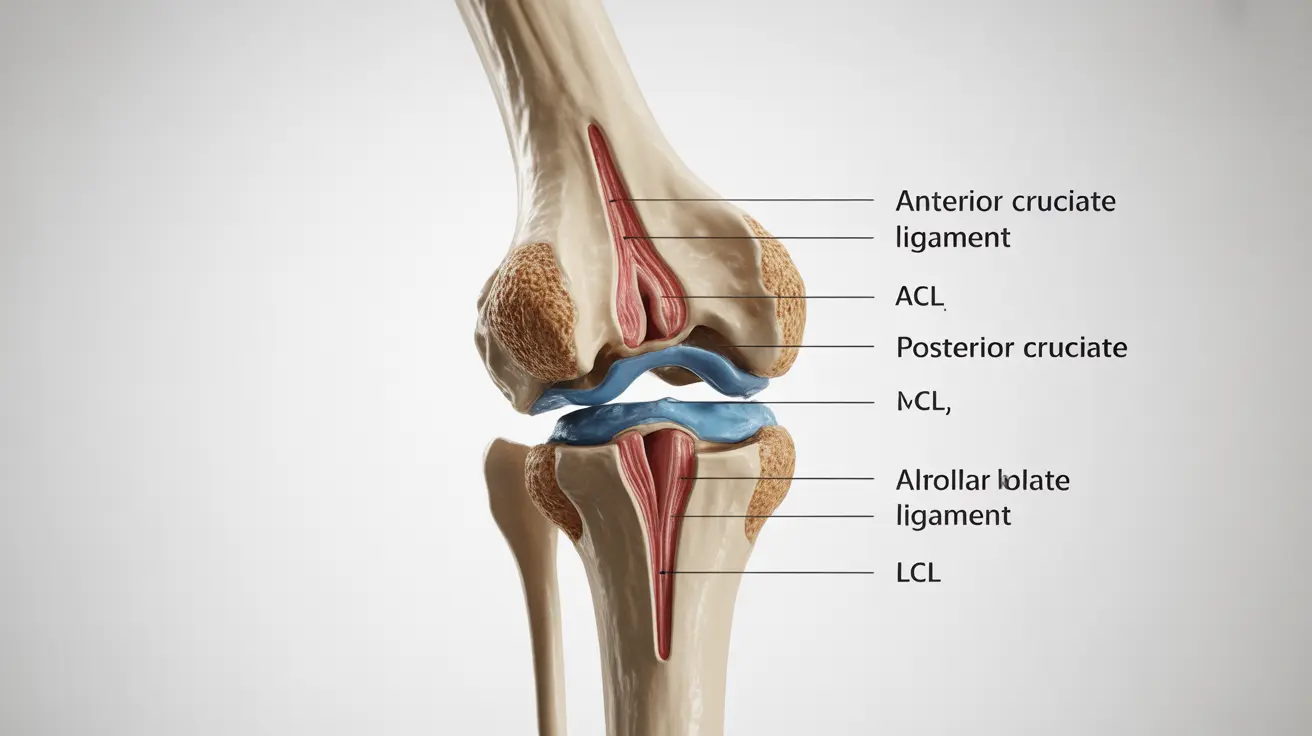
Four primary ligaments provide crucial stability to the knee joint:
- Anterior Cruciate Ligament (ACL): Controls forward movement of the tibia
- Posterior Cruciate Ligament (PCL): Prevents backward movement of the tibia
- Medial Collateral Ligament (MCL): Provides inner knee stability
- Lateral Collateral Ligament (LCL): Stabilizes the outer knee
These ligaments work together like internal stabilizing ropes, ensuring proper knee alignment during various movements and activities.
Cartilage and Menisci Components
The knee contains two types of cartilage structures that are vital for smooth joint function:
Articular Cartilage
This smooth, white tissue covers the ends of the femur, tibia, and the underside of the patella. It allows for nearly frictionless movement between the bones and helps absorb shock during physical activities.
Menisci
The medial and lateral menisci are C-shaped pieces of cartilage that act as natural shock absorbers between the femur and tibia. They help distribute weight evenly across the knee joint and improve overall joint stability.
Tendons and Muscles Around the Knee
Several key tendons and muscles support knee function:
- Quadriceps tendon: Connects the powerful quad muscles to the patella
- Patellar tendon: Links the patella to the tibia
- Hamstring tendons: Attach hamstring muscles to the tibia and fibula
- Iliotibial band: Runs along the outer thigh and knee
Supporting Soft Tissue Structures
The knee joint is enclosed in a synovial membrane that produces lubricating fluid. This membrane, along with various bursae (fluid-filled sacs), helps reduce friction between moving parts and provides necessary cushioning during movement.
Frequently Asked Questions
What are the main bones and ligaments labeled in a detailed knee anatomy diagram?
A detailed knee anatomy diagram shows three main bones: the femur (thighbone), tibia (shinbone), and patella (kneecap). The four major ligaments are the ACL, PCL, MCL, and LCL, each connecting different parts of the bones to provide stability.
How do the different ligaments and tendons in the knee contribute to its stability and movement?
Ligaments connect bones to bones, providing stability and preventing excessive movement in specific directions. The ACL and PCL control forward and backward motion, while the MCL and LCL provide side-to-side stability. Tendons connect muscles to bones, allowing for controlled movement and power generation.
What role do the menisci and cartilage play in knee joint function as seen in knee anatomy illustrations?
The menisci act as shock absorbers and weight distributors between the femur and tibia. Articular cartilage provides smooth surfaces for joint movement and helps absorb impact. Together, they protect the knee from wear and tear while enabling fluid motion.
How can understanding a labeled knee anatomy help identify common knee injuries like ACL or MCL tears?
Knowledge of knee anatomy helps identify injury locations and understand symptoms. For example, ACL tears often occur in the center of the knee and may cause instability during pivoting movements, while MCL injuries typically cause pain on the inner knee and may result from side impact.
What are the key soft tissue structures around the knee shown in anatomy diagrams and how do they support knee health?
Key soft tissue structures include the synovial membrane, which produces joint fluid for lubrication, bursae that reduce friction between moving parts, and the joint capsule that encloses the knee joint. These structures work together to maintain smooth movement and protect the joint from wear.


