


Lattice degeneration of the retina is a common eye condition that affects approximately 6-8% of the general population. This condition involves thinning and weakening of the peripheral retina, creating a lattice-like pattern of lesions that can potentially lead to serious complications if left unmonitored.
While many people with lattice degeneration never experience problems, understanding this condition is crucial because it can increase the risk of retinal tears and detachment, which may threaten vision if not addressed promptly.
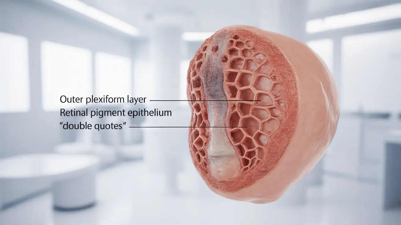
What is Lattice Degeneration?
Lattice degeneration appears as multiple small, oval-shaped areas of retinal thinning, typically in the peripheral retina. These areas often contain abnormal blood vessels and may develop small holes. The affected areas resemble a lattice or crisscross pattern, hence the name.
This condition usually develops in both eyes and tends to be more common in people who are nearsighted (myopic). The thinned areas can make the retina more vulnerable to tears or detachment, especially during sudden eye movements or trauma.
Signs and Symptoms
Most people with lattice degeneration don't experience any symptoms initially. However, certain warning signs may indicate complications:
- Sudden flashes of light (photopsia)
- New floaters or an increase in existing floaters
- A curtain-like shadow in peripheral vision
- Sudden decrease in vision
- Vision distortion or blurriness
These symptoms, particularly if they occur suddenly, may indicate a retinal tear or detachment and require immediate medical attention.
Diagnostic Process
Lattice degeneration is typically discovered during routine dilated eye examinations. Eye care professionals use various methods to diagnose the condition:
- Dilated fundus examination
- Indirect ophthalmoscopy
- Scleral depression
- Wide-field retinal imaging
These techniques allow doctors to carefully examine the peripheral retina and document any areas of concern for future monitoring.
Treatment Approaches
Treatment for lattice degeneration depends on several factors, including:
Preventive Treatment
In most cases, regular monitoring is sufficient if no complications are present. However, preventive treatment might be recommended for high-risk patients, including:
- Laser photocoagulation around weakened areas
- Cryotherapy (freezing treatment) in specific cases
- Regular follow-up examinations
Emergency Treatment
If a retinal tear or detachment occurs, immediate treatment is necessary and may include:
- Laser photocoagulation for tears
- Pneumatic retinopexy
- Scleral buckle surgery
- Vitrectomy
Risk Factors and Prevention
Several factors can increase the likelihood of developing lattice degeneration:
- Myopia (nearsightedness)
- Family history of retinal detachment
- Previous eye trauma
- Age (typically develops in young adults)
- Certain genetic conditions
Regular eye examinations are crucial for early detection and monitoring, especially for individuals with known risk factors.
Frequently Asked Questions
1. What is lattice degeneration of the retina and how common is it? Lattice degeneration is a condition where parts of the peripheral retina become thin and weak, affecting about 6-8% of people. It creates a characteristic lattice-like pattern and can increase the risk of retinal complications.
2. What symptoms might indicate a retinal tear or detachment from lattice degeneration? Warning signs include sudden flashes of light, new or increased floaters, a curtain-like shadow in vision, sudden vision decrease, or vision distortion. These symptoms require immediate medical attention.
3. How is lattice degeneration diagnosed during an eye exam? Diagnosis occurs through a dilated eye examination using indirect ophthalmoscopy, scleral depression, and sometimes wide-field retinal imaging. These tools allow thorough examination of the peripheral retina.
4. When and how is lattice degeneration treated, especially to prevent retinal detachment? Treatment depends on risk factors and complications. Most cases only require monitoring, but high-risk cases may need preventive laser treatment or cryotherapy. If complications occur, various surgical options are available.
5. What are the main risk factors for developing lattice degeneration of the retina? The main risk factors include myopia, family history of retinal detachment, previous eye trauma, young adult age, and certain genetic conditions. Regular eye examinations are essential for those with these risk factors.


