


A liver ultrasound is a valuable, non-invasive imaging procedure that helps healthcare providers examine the liver and surrounding organs. This diagnostic tool uses sound waves to create detailed images of your liver's structure, enabling doctors to detect various liver conditions and guide treatment decisions.
Understanding what to expect during a liver ultrasound and how it works can help ease any concerns you might have about the procedure. This guide will walk you through everything you need to know about liver ultrasounds, from preparation to results interpretation.
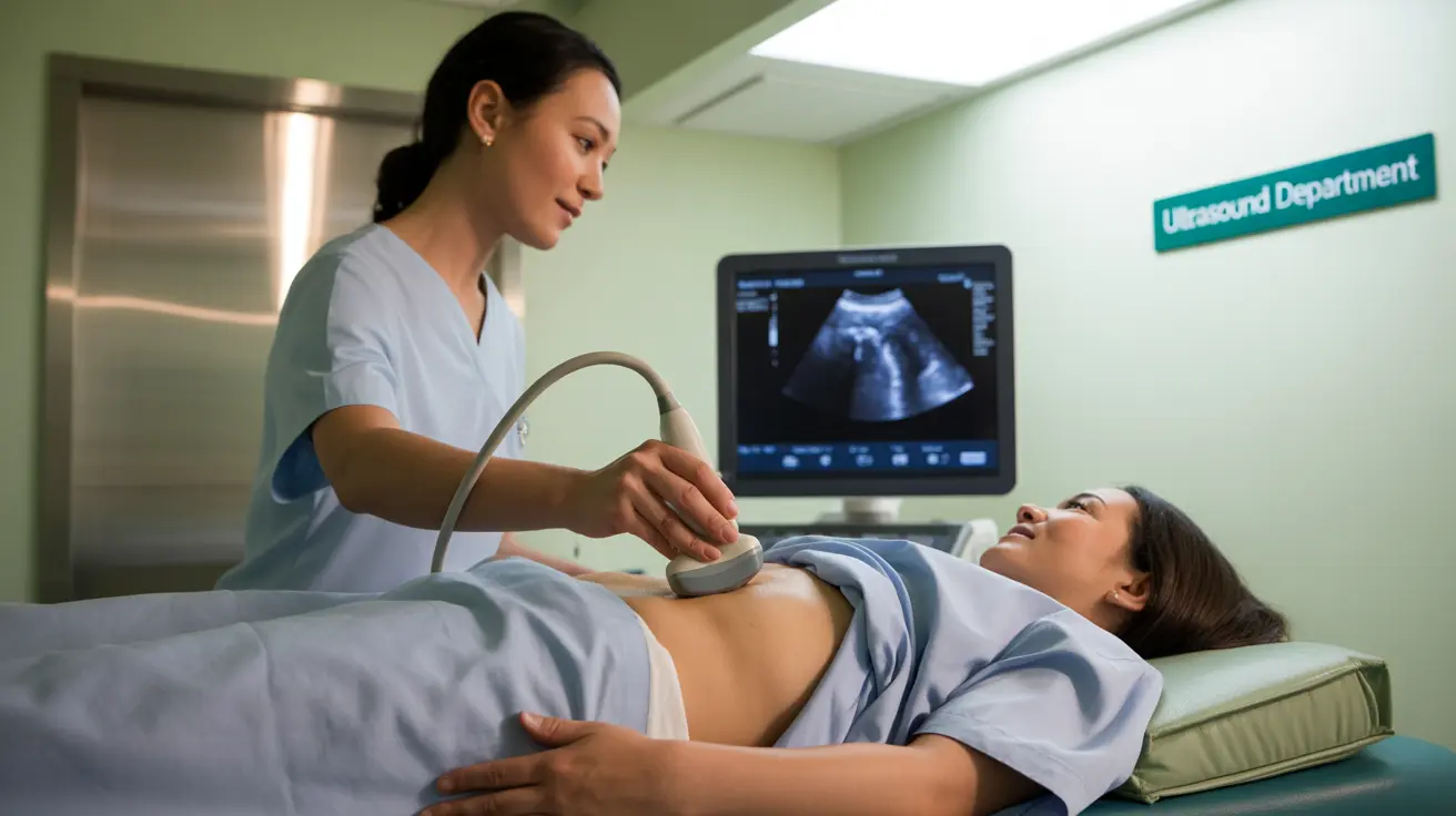
How Liver Ultrasound Works
During a liver ultrasound, a trained technician uses a handheld device called a transducer to send high-frequency sound waves through your body. These waves bounce off your internal organs and create echoes, which are converted into detailed images on a computer screen.
The procedure typically takes 20-30 minutes and is performed while you lie comfortably on an examination table. The technician will apply a special gel to your abdomen to help the sound waves travel more effectively between the transducer and your body.
Preparing for Your Liver Ultrasound
Proper preparation is crucial for obtaining accurate results from your liver ultrasound. Your healthcare provider may give you specific instructions, but general preparation guidelines typically include:
- Fasting for 6-8 hours before the examination
- Wearing loose, comfortable clothing
- Avoiding gas-producing foods for 24 hours before the procedure
- Taking medications as prescribed, unless instructed otherwise
Conditions Detected Through Liver Ultrasound
A liver ultrasound can identify various liver conditions and abnormalities, including:
Structural Changes
- Liver masses or tumors
- Cysts
- Changes in liver size
- Structural abnormalities
Common Liver Conditions
The ultrasound can help diagnose:
- Fatty liver disease
- Cirrhosis
- Liver cancer
- Blocked bile ducts
- Portal hypertension
Advanced Liver Imaging: Elastography
Liver elastography is a specialized type of ultrasound that measures liver stiffness to assess the presence and severity of liver fibrosis. This technique provides additional information about liver health that may not be visible on standard ultrasound images.
The procedure is particularly useful for:
- Monitoring liver disease progression
- Evaluating the need for liver biopsy
- Assessing treatment effectiveness
Safety and Risk Considerations
Liver ultrasound is considered one of the safest diagnostic imaging procedures available. It doesn't use radiation, making it suitable for repeated examinations when necessary. The procedure is:
- Pain-free and non-invasive
- Safe for pregnant women
- Free from radiation exposure
- Suitable for all age groups
Frequently Asked Questions
What is a liver ultrasound and how is the procedure performed?
A liver ultrasound is a diagnostic imaging procedure that uses sound waves to create pictures of your liver. During the procedure, a technician moves a transducer device across your abdomen while you lie on an examination table. The entire process typically takes 20-30 minutes and is painless.
How should I prepare for a liver ultrasound to ensure accurate results?
To prepare for a liver ultrasound, you should fast for 6-8 hours before the examination, wear comfortable clothing, and avoid gas-producing foods for 24 hours prior. Follow any specific instructions provided by your healthcare provider regarding medications.
What conditions can a liver ultrasound detect, such as fatty liver or cirrhosis?
A liver ultrasound can detect various conditions including fatty liver disease, cirrhosis, liver tumors, cysts, blocked bile ducts, and structural abnormalities. It can also help identify changes in liver size and texture that might indicate underlying health issues.
Is a liver ultrasound safe, and are there any risks or side effects?
Liver ultrasounds are extremely safe with no known risks or side effects. The procedure is non-invasive, doesn't use radiation, and is safe for all patients, including pregnant women and children.
How does liver elastography differ from a standard liver ultrasound and what does it measure?
Liver elastography is a specialized ultrasound technique that measures liver stiffness to assess fibrosis levels. Unlike standard ultrasound, which provides images of liver structure, elastography specifically measures tissue elasticity to evaluate liver scarring and disease progression without the need for invasive biopsies.


