


When receiving mammogram results, one of the most common concerns is understanding the difference between breast cysts and potentially cancerous masses. This distinction is crucial for proper diagnosis and peace of mind, as these two findings can appear similar on imaging but have vastly different implications for health and treatment.
Learning to understand how medical professionals differentiate between cysts and cancer through various imaging techniques can help you better navigate your breast health journey and understand what to expect during the diagnostic process.
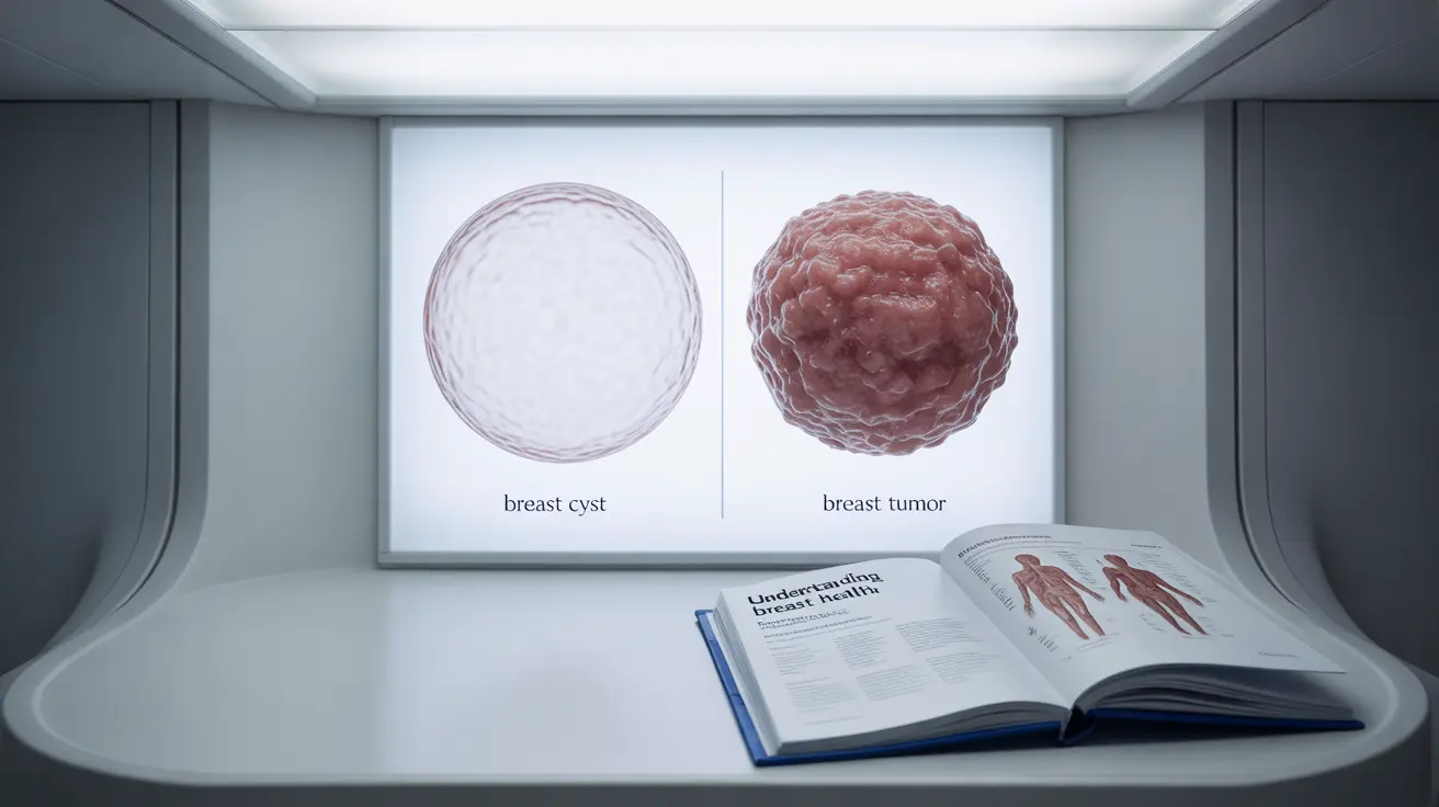
Key Characteristics of Breast Cysts and Tumors
Breast cysts and tumors have distinct characteristics that help medical professionals identify them through imaging studies:
Breast Cyst Features
- Fluid-filled sacs with clear boundaries
- Usually round or oval in shape
- Often appear dark and uniform on ultrasound
- May change size during menstrual cycles
- Generally smooth and mobile when touched
Tumor Characteristics
- Solid masses with varying density
- May have irregular shapes or borders
- Usually appear as white or light areas on mammograms
- Typically maintain consistent size
- Often feel firm and less mobile
Imaging Technologies Used for Diagnosis
Mammogram Capabilities
Mammograms serve as the primary screening tool, creating detailed X-ray images of breast tissue. They can reveal calcifications, density changes, and masses, but sometimes require additional imaging for definitive diagnosis.
Ultrasound Assessment
Ultrasound technology is particularly effective at distinguishing fluid-filled cysts from solid masses. It uses sound waves to create detailed images and can show whether a mass is solid or fluid-filled with high accuracy.
When Additional Testing Is Needed
Sometimes, initial imaging results may be inconclusive, requiring additional diagnostic procedures:
Further Imaging
- Magnetic Resonance Imaging (MRI)
- 3D mammography (tomosynthesis)
- Contrast-enhanced mammography
Biopsy Considerations
If imaging cannot definitively determine whether a mass is benign or potentially cancerous, a biopsy may be necessary. This involves taking a small sample of tissue for laboratory analysis.
Follow-up Care and Monitoring
The type of follow-up care recommended depends on the initial findings and risk factors. Regular monitoring may be advised for certain types of benign findings, while suspicious masses will require more immediate attention and possibly intervention.
Frequently Asked Questions
What is the difference between a breast cyst and a tumor found on a mammogram, and how do doctors tell them apart using imaging tests? Doctors distinguish between cysts and tumors using various imaging characteristics. Cysts typically appear as well-defined, fluid-filled sacs on ultrasound and mammogram, while tumors are solid masses that may have irregular borders and different density patterns.
Can a mammogram or ultrasound reliably distinguish between a harmless breast cyst and cancer, and what happens if the result is unclear? While mammograms and ultrasounds are generally reliable in distinguishing cysts from solid masses, sometimes results may be unclear. In these cases, additional imaging tests or a biopsy may be necessary for definitive diagnosis.
What symptoms should make me worry that a breast lump might be cancer and not just a cyst? Concerning symptoms include hardness of the mass, lack of mobility, irregular borders, skin changes, nipple discharge or changes, and persistence without size fluctuation. Any new or changing breast symptoms should be evaluated by a healthcare provider.
If a mammogram shows a breast cyst, is there any risk it could be cancerous or turn into cancer later? Simple breast cysts are typically benign and do not increase cancer risk. However, complex cysts or those with solid components may require additional evaluation to ensure they don't contain cancerous elements.
What follow-up tests or treatments are usually recommended after finding a cyst versus a solid mass on breast imaging? Simple cysts typically require no treatment unless they cause discomfort. They may be monitored with regular screening mammograms. Solid masses often require additional imaging, possible biopsy, and more frequent follow-up based on their characteristics and risk assessment.


