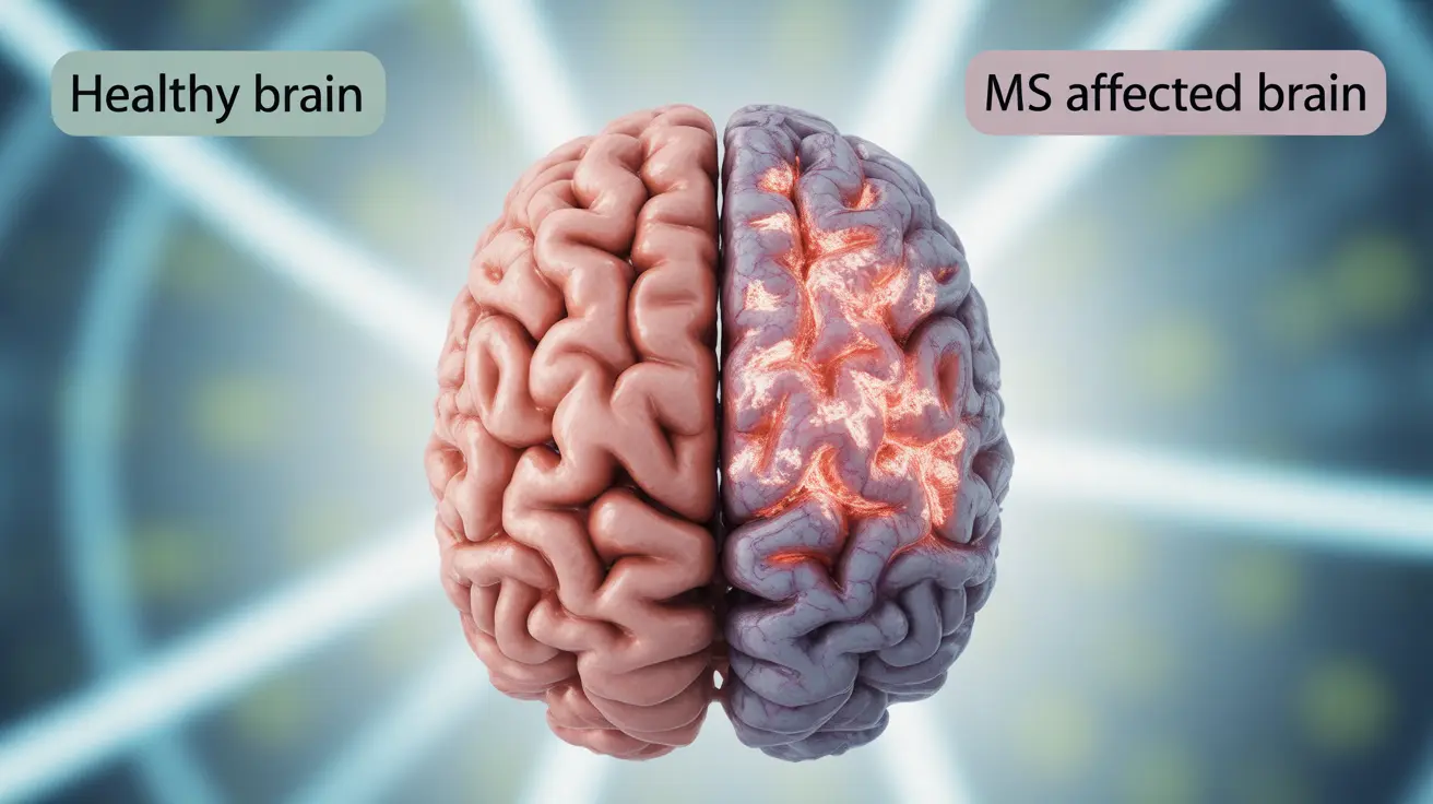
Multiple sclerosis (MS) causes distinct changes in the brain that differentiate it from a healthy brain. Understanding these differences is crucial for both medical professionals and patients, as it helps explain the varied symptoms and progression of MS. This comprehensive guide explores how MS affects brain structure, function, and what these changes mean for people living with the condition.
Visible Changes in Brain Structure
When comparing an MS brain to a normal brain through imaging techniques, several key structural differences become apparent. The most notable changes include lesions or plaques in the white matter, which appear as bright spots on MRI scans. These lesions represent areas where the protective myelin coating around nerve fibers has been damaged or destroyed by the immune system.
Brain volume changes also become evident as MS progresses. People with MS may experience more rapid brain atrophy compared to those without the condition, particularly affecting areas responsible for cognitive function and motor control.
Impact on Myelin and Nerve Signaling
The primary distinction between an MS brain and a normal brain lies in how nerve signals are transmitted. In a healthy brain, myelin sheaths ensure rapid and efficient signal transmission between neurons. However, in MS:
- Damaged myelin slows or disrupts nerve signal transmission
- Communication between different brain regions becomes less efficient
- The body attempts to repair myelin, but often incompletely
- Nerve fibers themselves may eventually become damaged
Common Symptoms Related to Brain Changes
The structural and functional differences between an MS brain and a normal brain manifest in various symptoms:
Cognitive Effects
- Memory problems
- Difficulty concentrating
- Slower information processing
- Challenges with complex problem-solving
Physical Manifestations
- Balance difficulties
- Coordination problems
- Vision changes
- Muscle weakness or spasticity
Brain Atrophy and Disease Progression
Brain atrophy in MS occurs at a faster rate than normal age-related brain volume loss. Modern imaging techniques can detect these changes early, helping doctors monitor disease progression and adjust treatment strategies accordingly. Understanding the rate and pattern of brain atrophy helps predict disease course and potential disability risks.
Role of Medical Imaging
Medical imaging plays a crucial role in distinguishing an MS brain from a normal brain. Different techniques provide valuable information:
MRI Scanning
- Shows active inflammation
- Reveals new and old lesions
- Monitors brain volume changes
- Helps track disease progression
Advanced Imaging Technologies
- Functional MRI shows brain activity patterns
- Diffusion tensor imaging reveals white matter integrity
- Spectroscopy examines brain tissue metabolism
Frequently Asked Questions
What are the main structural differences between an MS brain and a normal brain on MRI scans? The main differences include visible lesions or plaques in the white matter, areas of inflammation, and potentially increased brain atrophy. These appear as bright or dark spots on MRI scans, depending on the scanning technique used.
How does damage to myelin in the brain affect nerve signals in people with multiple sclerosis? Myelin damage slows or disrupts nerve signal transmission, leading to delayed or interrupted communication between different parts of the brain. This disruption results in various neurological symptoms and can affect both motor and cognitive functions.
What symptoms are caused by the changes that occur in the brain due to multiple sclerosis? Brain changes in MS can cause a wide range of symptoms, including cognitive difficulties (memory problems, concentration issues), physical symptoms (balance problems, muscle weakness), vision changes, and fatigue. The specific symptoms vary depending on which areas of the brain are affected.
Can brain atrophy in multiple sclerosis patients be detected early, and what does it indicate? Yes, modern imaging techniques can detect brain atrophy early in the disease course. Accelerated brain atrophy often indicates disease progression and may correlate with future disability risks. Early detection helps guide treatment decisions.
How do MRI and other imaging techniques help doctors diagnose and monitor multiple sclerosis progression? Imaging techniques, particularly MRI, help doctors identify new lesions, monitor disease activity, track brain volume changes, and assess treatment effectiveness. These tools are essential for both initial diagnosis and ongoing disease management.


