


Brain MRI (Magnetic Resonance Imaging) plays a crucial role in understanding and evaluating Parkinson's disease, though its relationship with diagnosis is complex and often misunderstood. While MRI cannot definitively diagnose Parkinson's disease on its own, it provides valuable insights into brain changes and helps medical professionals rule out other conditions.
This comprehensive guide explores how brain MRI scans differ between individuals with Parkinson's disease and those with healthy brains, helping you understand the role of this important diagnostic tool in neurological care.
Key MRI Differences in Parkinson's Disease
When comparing brain MRI scans of Parkinson's patients to those of healthy individuals, several distinctive features may be observed:
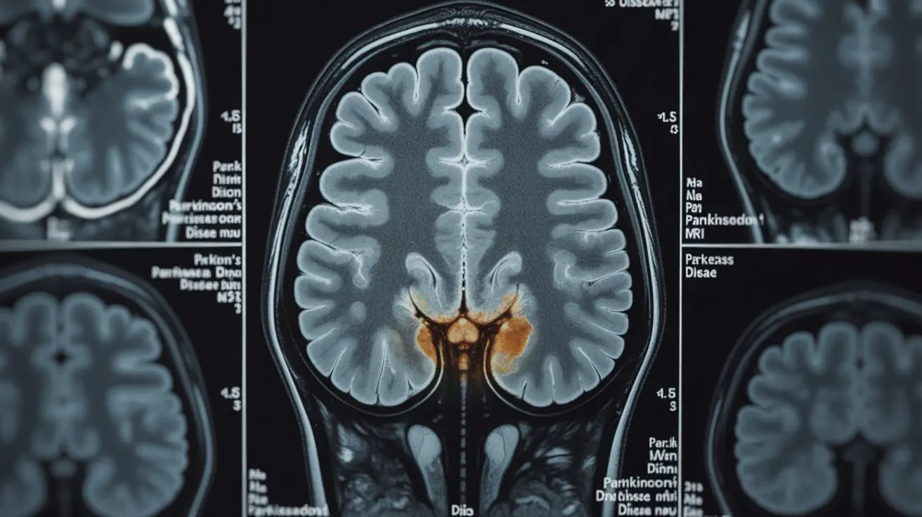
Changes in the Substantia Nigra
The substantia nigra, a critical brain region for movement control, typically shows reduced volume and altered signal intensity in Parkinson's patients. Advanced MRI techniques can detect the loss of dopamine-producing neurons in this area, though these changes may not be visible on standard MRI scans.
Brain Volume and Structure
Parkinson's disease can lead to various structural changes visible on MRI, including:
- Reduced gray matter volume in specific brain regions
- Changes in white matter integrity
- Possible enlargement of the ventricles
- Alterations in cortical thickness
Advanced MRI Techniques for Parkinson's Assessment
Diffusion Tensor Imaging (DTI)
DTI helps visualize the brain's white matter tracts, revealing potential disruptions in neural connectivity that may occur in Parkinson's disease. This specialized technique can detect subtle changes before they become apparent on conventional MRI scans.
Functional MRI (fMRI)
fMRI examines brain activity patterns during rest or specific tasks, helping researchers understand how Parkinson's affects neural circuits and potentially identifying early functional changes before structural alterations appear.
MRI's Role in Differential Diagnosis
While MRI cannot definitively diagnose Parkinson's disease, it serves several crucial purposes in the diagnostic process:
- Ruling out other neurological conditions
- Identifying vascular problems that might mimic Parkinson's symptoms
- Monitoring disease progression
- Supporting clinical diagnosis when combined with other diagnostic tools
Limitations of MRI in Parkinson's Diagnosis
Understanding the limitations of MRI in Parkinson's diagnosis is essential:
- Early-stage changes may not be visible on conventional MRI
- Normal MRI results don't rule out Parkinson's disease
- Some changes visible on MRI can be associated with normal aging
- Individual variation can make interpretation challenging
Frequently Asked Questions
How does a Parkinson's brain MRI differ from a normal brain MRI?
A Parkinson's brain MRI may show reduced volume in the substantia nigra, changes in brain structure, and alterations in white matter integrity. However, these changes aren't always visible on standard MRI scans, especially in early stages of the disease.
Can an MRI detect early signs of Parkinson's disease before symptoms appear?
Standard MRI typically cannot detect Parkinson's disease before symptoms appear. However, advanced techniques like DTI and fMRI may help identify subtle brain changes before clinical symptoms become evident, though this is primarily used in research settings.
What specific MRI features indicate Parkinson's disease compared to a healthy brain?
Key MRI features may include reduced substantia nigra volume, changes in brain structure, altered white matter integrity, and possible ventricle enlargement. However, these changes aren't universal or specific to Parkinson's disease.
How is MRI used to distinguish Parkinson's disease from other similar neurological conditions?
MRI helps differentiate Parkinson's from other conditions by revealing specific patterns of brain change and ruling out other causes of symptoms, such as strokes, tumors, or other neurodegenerative disorders.
Why can't an MRI alone definitively diagnose Parkinson's disease?
MRI cannot definitively diagnose Parkinson's because the disease's early changes may not be visible on scans, and some visible changes can overlap with normal aging or other conditions. Diagnosis requires a combination of clinical evaluation, patient history, and multiple diagnostic tools.


