


Understanding how pelvic inflammatory disease (PID) appears on ultrasound is crucial for both healthcare providers and patients. While ultrasound serves as an important diagnostic tool, its effectiveness can vary depending on the stage and severity of the condition. This guide explores the relationship between PID and ultrasound imaging, helping you understand what to expect during the diagnostic process.
Understanding Ultrasound in PID Diagnosis
Ultrasound imaging plays a vital role in evaluating pelvic inflammatory disease, though it's important to note that it's just one of several diagnostic tools available. Healthcare providers typically use ultrasound in conjunction with other methods to confirm a PID diagnosis and assess its severity.
Types of Ultrasound Used for PID Detection
Transvaginal Ultrasound
This method provides detailed images of the reproductive organs by placing a special ultrasound probe in the vagina. It offers clearer visualization of potential PID-related changes compared to traditional abdominal ultrasound.
Transabdominal Ultrasound
This conventional ultrasound method can help identify larger abnormalities and is often used alongside transvaginal ultrasound for a complete evaluation.
What Ultrasound Can Reveal About PID
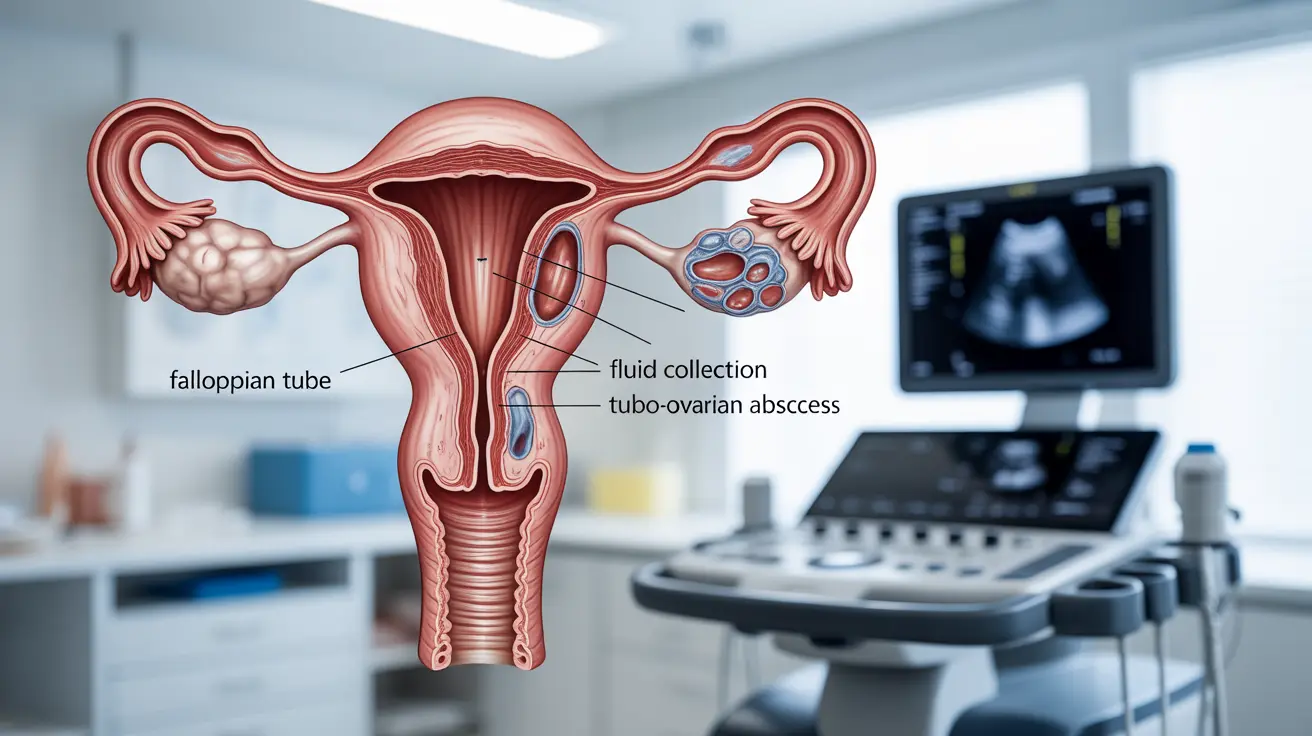
Ultrasound can detect several indicators of pelvic inflammatory disease, particularly in more advanced cases. These may include:
- Thickened fallopian tubes
- Fluid collections in the pelvic area
- Tubo-ovarian abscesses
- Adhesions or scarring
- Enhanced vascularity in affected areas
Limitations of Ultrasound in PID Diagnosis
While ultrasound is valuable, it has certain limitations in detecting PID. Early-stage cases may not show clear signs on ultrasound, and some findings can be subtle or nonspecific. This is why healthcare providers often rely on multiple diagnostic approaches.
When Doctors Order Ultrasound Studies
Healthcare providers typically recommend ultrasound imaging when:
- Symptoms are severe or persistent
- There's uncertainty about the diagnosis
- Complications are suspected
- Treatment isn't producing expected results
- Regular monitoring of known PID is needed
Frequently Asked Questions
Does pelvic inflammatory disease always show up on an ultrasound scan?
No, PID doesn't always show up on ultrasound, especially in early stages. Early PID may have minimal or no visible changes on ultrasound, which is why doctors also rely on clinical symptoms and other diagnostic methods.
What ultrasound signs indicate advanced pelvic inflammatory disease?
Advanced PID typically shows thickened fallopian tubes, fluid collections, tubo-ovarian abscesses, and increased blood flow in affected areas. These findings are more readily visible on ultrasound compared to early-stage disease.
How effective is transvaginal ultrasound in diagnosing early-stage PID?
Transvaginal ultrasound has limited effectiveness in diagnosing early-stage PID. While it provides detailed images of the reproductive organs, subtle early changes may not be visible. It's more useful for detecting advanced disease or complications.
When do doctors recommend imaging tests for pelvic inflammatory disease?
Doctors typically recommend imaging tests when symptoms are severe, the diagnosis is unclear, complications are suspected, or when monitoring treatment response. They may also order ultrasounds for patients who aren't improving with standard treatment.
Can ultrasound detect complications like abscesses or scarring caused by pelvic inflammatory disease?
Yes, ultrasound is particularly effective at detecting PID complications such as abscesses, fluid collections, and scarring. These complications are usually more visible on ultrasound than early-stage inflammation.
While ultrasound is a valuable tool in diagnosing and monitoring PID, it's important to remember that it's just one part of a comprehensive diagnostic approach. Always consult with healthcare providers for proper evaluation and treatment of potential PID symptoms.


