


Psoriatic arthritis affects millions of people worldwide, causing joint inflammation, pain, and potential long-term damage. When doctors suspect this condition, imaging studies like X-rays play a crucial role in diagnosis and ongoing monitoring. Understanding what a psoriatic arthritis X-ray can reveal helps patients and healthcare providers make informed decisions about treatment and care.
X-ray imaging remains one of the most accessible and valuable tools for evaluating joint changes associated with psoriatic arthritis. While other imaging methods may be necessary for early detection, X-rays provide essential information about bone structure, joint space, and characteristic changes that distinguish psoriatic arthritis from other inflammatory conditions.
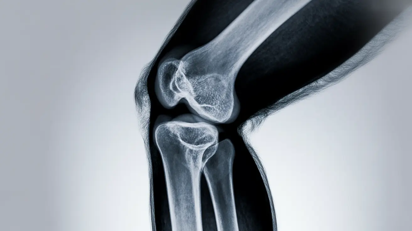
Distinctive X-Ray Changes in Psoriatic Arthritis
A psoriatic arthritis X-ray reveals several unique features that help differentiate this condition from rheumatoid arthritis and other inflammatory joint diseases. These characteristic changes develop over time as the condition progresses, creating a distinctive pattern that experienced radiologists and rheumatologists can recognize.
One of the most notable findings on X-ray imaging is asymmetric joint involvement. Unlike rheumatoid arthritis, which typically affects joints symmetrically on both sides of the body, psoriatic arthritis often impacts joints in an irregular pattern. This asymmetry appears clearly on X-ray images and serves as an important diagnostic clue.
Joint space narrowing occurs as cartilage deteriorates, but in psoriatic arthritis, this narrowing may be accompanied by bone formation at tendon and ligament attachment points. These areas, called entheses, show characteristic changes that distinguish psoriatic arthritis from other conditions on X-ray examination.
Bone erosions in psoriatic arthritis often have a "fluffy" or irregular appearance, contrasting with the clean, well-defined erosions typically seen in rheumatoid arthritis. Additionally, X-rays may show new bone formation alongside destructive changes, creating a mixed pattern of destruction and repair that is characteristic of this condition.
Early Detection Limitations and Alternative Imaging
While X-rays are valuable for diagnosing established psoriatic arthritis, they have significant limitations in detecting early disease. Initial joint damage may not be visible on standard X-ray images for months or even years after symptoms begin. This delay in visible changes means that early diagnosis often requires additional imaging methods.
Magnetic resonance imaging (MRI) and ultrasound are more sensitive tools for detecting early inflammatory changes in psoriatic arthritis. These imaging methods can identify soft tissue inflammation, early cartilage damage, and bone marrow changes before they become apparent on X-rays. Many rheumatologists now use these advanced imaging techniques when X-rays appear normal but clinical suspicion remains high.
The combination of clinical examination, laboratory tests, and multiple imaging methods provides the most comprehensive approach to early diagnosis. While X-rays may not show changes initially, they become increasingly valuable as the disease progresses and structural damage develops.
The Pencil-in-Cup Deformity: A Hallmark Finding
The pencil-in-cup deformity represents one of the most characteristic X-ray findings in advanced psoriatic arthritis. This distinctive appearance occurs when severe bone erosion creates a cup-shaped depression in one bone end, while the opposing bone develops a pointed, pencil-like shape that fits into the cup.
This deformity most commonly affects the small joints of the hands and feet, particularly the distal interphalangeal joints. The pencil-in-cup appearance results from the unique pattern of bone destruction and remodeling that occurs in psoriatic arthritis, where erosive changes are accompanied by new bone formation.
When radiologists identify pencil-in-cup deformities on X-ray images, it strongly suggests psoriatic arthritis rather than other inflammatory conditions. This finding is so characteristic that it often confirms the diagnosis even when other features are less clear. However, pencil-in-cup deformities typically indicate advanced disease, emphasizing the importance of early diagnosis and treatment.
Monitoring Disease Progression with X-Rays
Regular X-ray monitoring plays an essential role in managing psoriatic arthritis over time. The frequency of imaging depends on various factors, including disease activity, treatment response, and individual risk factors for progression. Most rheumatologists recommend baseline X-rays at diagnosis, followed by periodic imaging to assess treatment effectiveness and detect new damage.
For patients with active disease or those starting new treatments, X-rays may be repeated every 6 to 12 months initially. Once the condition stabilizes or goes into remission, the interval between X-rays may extend to 1 to 2 years or longer. The specific monitoring schedule should always be individualized based on clinical circumstances and physician recommendations.
Comparing serial X-rays allows healthcare providers to track disease progression objectively. New erosions, increasing joint space narrowing, or developing deformities indicate that current treatment may need adjustment. Conversely, stable X-ray findings suggest that therapy is effectively controlling the inflammatory process.
Safety Considerations for Repeated X-Ray Exposure
While X-rays are generally safe, repeated exposure does involve cumulative radiation risk that patients and providers should consider. The radiation dose from a typical joint X-ray is relatively low, equivalent to a few days of natural background radiation exposure. However, multiple X-rays over many years can add up to more significant exposure.
Modern X-ray equipment and techniques minimize radiation exposure while maintaining image quality. Digital radiography systems, now standard in most facilities, use lower radiation doses than older film-based systems. Additionally, focused imaging of specific joints rather than whole-body surveys reduces unnecessary exposure.
Healthcare providers carefully balance the benefits of monitoring disease progression against radiation risks. For most patients with psoriatic arthritis, the valuable information gained from periodic X-rays far outweighs the small associated radiation risk. Pregnant women require special consideration, and alternative imaging methods may be preferred when possible.
Frequently Asked Questions
What changes can psoriatic arthritis show on an X-ray, and how are these different from other types of arthritis?
Psoriatic arthritis X-rays show distinctive features including asymmetric joint involvement, irregular "fluffy" bone erosions, new bone formation at tendon attachment sites, and joint space narrowing. Unlike rheumatoid arthritis, which creates symmetrical, well-defined erosions, psoriatic arthritis produces mixed destructive and constructive bone changes in an asymmetrical pattern throughout the body.
Can an X-ray detect early signs of psoriatic arthritis, or is another imaging test needed at first?
X-rays have limited ability to detect early psoriatic arthritis signs, as structural changes may not appear for months or years after symptom onset. MRI and ultrasound are more sensitive for early detection, identifying soft tissue inflammation, cartilage damage, and bone marrow changes before they become visible on X-rays. Early diagnosis often requires combining clinical assessment with advanced imaging techniques.
What is a pencil-in-cup deformity on an X-ray, and why is it important for diagnosing psoriatic arthritis?
A pencil-in-cup deformity appears when severe bone erosion creates a cup-shaped depression in one bone end while the opposing bone develops a pointed, pencil-like shape. This characteristic finding, commonly seen in finger and toe joints, is highly specific for psoriatic arthritis and helps distinguish it from other inflammatory conditions. However, it typically indicates advanced disease requiring prompt treatment.
How often should people with psoriatic arthritis get X-rays to check for joint damage or disease progression?
X-ray frequency varies based on disease activity and treatment response. Active disease may require imaging every 6-12 months initially, while stable or remission cases may need X-rays every 1-2 years or longer. Baseline X-rays at diagnosis establish a comparison point for future monitoring. Your rheumatologist will determine the appropriate schedule based on your individual circumstances and treatment plan.
Are there any risks or side effects from repeated X-rays when monitoring psoriatic arthritis?
Repeated X-rays involve cumulative radiation exposure, though individual joint X-rays produce relatively low radiation doses equivalent to a few days of natural background exposure. Modern digital equipment minimizes radiation while maintaining image quality. For most patients, the benefits of monitoring disease progression outweigh the small radiation risks, but healthcare providers carefully balance these considerations when planning imaging schedules.


