


When discovering a soft tissue mass, understanding the differences between sarcomas and lipomas through ultrasound imaging is crucial for proper diagnosis and treatment planning. Ultrasound serves as a valuable initial screening tool, offering important insights into the characteristics of these soft tissue masses, though it's important to understand both its capabilities and limitations.
This comprehensive guide explores how medical professionals use ultrasound to differentiate between sarcomas and lipomas, discussing key imaging features, diagnostic accuracy, and when additional testing may be necessary.
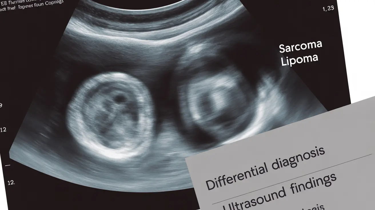
Key Ultrasound Characteristics in Differentiation
Ultrasound imaging reveals several distinct features that help healthcare providers distinguish between sarcomas and lipomas:
Appearance and Echogenicity
Lipomas typically appear as well-defined, oval-shaped masses with homogeneous internal echoes. They usually show uniform, low-level echogenicity and are compressible under gentle pressure. In contrast, sarcomas often display irregular borders, heterogeneous internal echoes, and may show areas of varying echogenicity.
Size and Depth Considerations
While size alone isn't definitive, sarcomas tend to be larger and grow more rapidly than lipomas. Ultrasound can effectively measure the mass's dimensions and depth within the tissue, which are important factors in risk assessment. Masses larger than 5 centimeters or located in deeper tissue layers warrant closer investigation.
Advanced Ultrasound Assessment Techniques
Doppler Evaluation
Color Doppler ultrasound provides crucial information about blood flow within the mass. Lipomas typically show minimal to no internal vascularity. Sarcomas, however, often demonstrate increased internal blood flow and irregular vascular patterns, which can be a significant indicator of malignancy.
Elastography Findings
Modern ultrasound machines equipped with elastography can assess tissue stiffness. Lipomas generally appear soft and elastic, while sarcomas tend to be harder and less compressible. This additional information can help strengthen the initial assessment.
Diagnostic Accuracy and Limitations
While ultrasound is an excellent initial imaging tool, it has certain limitations. The accuracy of ultrasound in distinguishing between sarcomas and lipomas varies depending on several factors, including the operator's expertise, the quality of the equipment, and the specific characteristics of the mass.
When Additional Imaging Is Necessary
Healthcare providers often recommend additional imaging studies when ultrasound findings are inconclusive or suggest concerning features. Common scenarios include:
- Deep-seated masses
- Lesions larger than 5 centimeters
- Masses with irregular borders or increased vascularity
- Rapidly growing or painful masses
Frequently Asked Questions
What ultrasound features help distinguish sarcoma from lipoma in soft tissue lumps?
Key ultrasound features that help distinguish sarcomas from lipomas include border definition, internal echo patterns, and compressibility. Lipomas typically show well-defined borders and uniform echogenicity, while sarcomas often have irregular borders and heterogeneous internal echoes.
How accurate is ultrasound in diagnosing soft tissue sarcoma compared to lipoma?
While ultrasound is highly useful as an initial screening tool, its accuracy varies. It's most accurate when combined with clinical history and physical examination. The sensitivity and specificity improve significantly when performed by experienced sonographers using high-quality equipment.
When should additional imaging like MRI or biopsy be done after an ultrasound shows a suspicious lump?
Additional imaging should be pursued when ultrasound reveals concerning features such as large size (>5cm), deep location, irregular borders, heterogeneous appearance, or significant vascularity. MRI or biopsy may also be warranted if the mass is growing rapidly or causing pain.
What does increased vascularity on Doppler ultrasound indicate in differentiating sarcoma from lipoma?
Increased vascularity on Doppler ultrasound often suggests potential malignancy, as sarcomas typically show greater internal blood flow and irregular vascular patterns compared to lipomas, which usually demonstrate minimal to no internal vascularity.
Can ultrasound alone definitively diagnose lipoma or sarcoma, or are further tests always needed?
While ultrasound provides valuable initial information, it cannot definitively diagnose sarcoma or lipoma on its own. Further testing, such as MRI or biopsy, is often necessary for definitive diagnosis, especially when suspicious features are present on ultrasound.


