


When doctors suspect a spinal tumor, medical imaging plays a crucial role in diagnosis, treatment planning, and monitoring. Understanding what spinal tumor pictures reveal through various imaging techniques can help patients better comprehend their condition and treatment journey.
This comprehensive guide explores how medical professionals use different imaging methods to identify and assess spinal tumors, what these images typically show, and what patients can expect during the diagnostic process.
Understanding Spinal Tumor Imaging Techniques
Medical professionals use several sophisticated imaging technologies to detect and evaluate spinal tumors. Each method provides unique and valuable information about the tumor's characteristics.
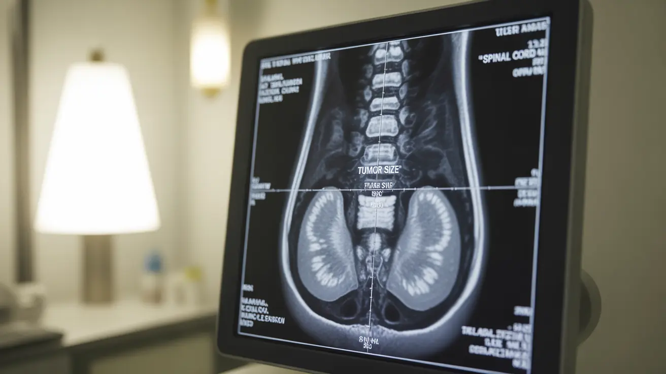
Magnetic Resonance Imaging (MRI)
MRI scans are the gold standard for spinal tumor detection. These detailed images show:
- Tumor size and location within the spine
- Relationship to surrounding tissues
- Extent of spinal cord compression
- Tumor characteristics that help determine if it's benign or malignant
Computed Tomography (CT) Scans
CT scans offer complementary information to MRI scans, particularly showing:
- Bone involvement and destruction
- Calcifications within tumors
- Detailed cross-sectional views of the spine
- Guide for surgical planning
Recognizing Key Features in Spinal Tumor Images
Medical professionals look for specific characteristics when examining spinal tumor pictures:
- Abnormal masses or lesions
- Changes in bone density
- Compression of nerve roots
- Alterations in spinal alignment
- Signs of surrounding tissue involvement
Common Signs and Symptoms
While imaging is essential for diagnosis, understanding the symptoms that prompt imaging is equally important:
- Back pain that worsens at night
- Unexplained neurological symptoms
- Progressive muscle weakness
- Difficulty walking
- Loss of bladder or bowel control
The Diagnostic Process
Diagnosis typically follows a structured approach:
- Initial physical examination
- Neurological assessment
- Multiple imaging studies
- Possible biopsy for definitive diagnosis
- Regular monitoring through follow-up scans
Treatment Planning Through Imaging
Spinal tumor pictures guide treatment decisions by helping doctors:
- Determine the best surgical approach
- Plan radiation therapy
- Monitor treatment response
- Assess for potential complications
- Track tumor progression or regression
Frequently Asked Questions
What do spinal tumor pictures from MRI and CT scans typically show? MRI and CT scans show detailed images of the tumor's size, location, and characteristics. MRI excels at showing soft tissue involvement, while CT scans better demonstrate bone involvement and can reveal calcifications within tumors.
What are the common symptoms that might indicate a spinal tumor? Common symptoms include persistent back pain (especially at night), neurological symptoms like numbness or weakness, difficulty walking, and changes in bladder or bowel function. These symptoms often prompt doctors to order imaging studies.
How do doctors use imaging like MRI to diagnose spinal tumors? Doctors use MRI as the primary imaging tool for diagnosing spinal tumors. They analyze multiple views and sequences to assess the tumor's characteristics, location, and impact on surrounding tissues. This information helps determine the tumor type and guides treatment planning.
What treatment options are available for spinal tumors once detected? Treatment options may include surgery, radiation therapy, chemotherapy, or a combination approach. The choice depends on the tumor's characteristics seen in imaging studies, its location, and whether it's primary or metastatic.
When should I see a doctor if I have back pain that might be related to a spinal tumor? Seek medical attention if you experience persistent back pain that worsens at night, doesn't improve with conservative treatment, or is accompanied by neurological symptoms like weakness, numbness, or changes in bladder/bowel function.


