


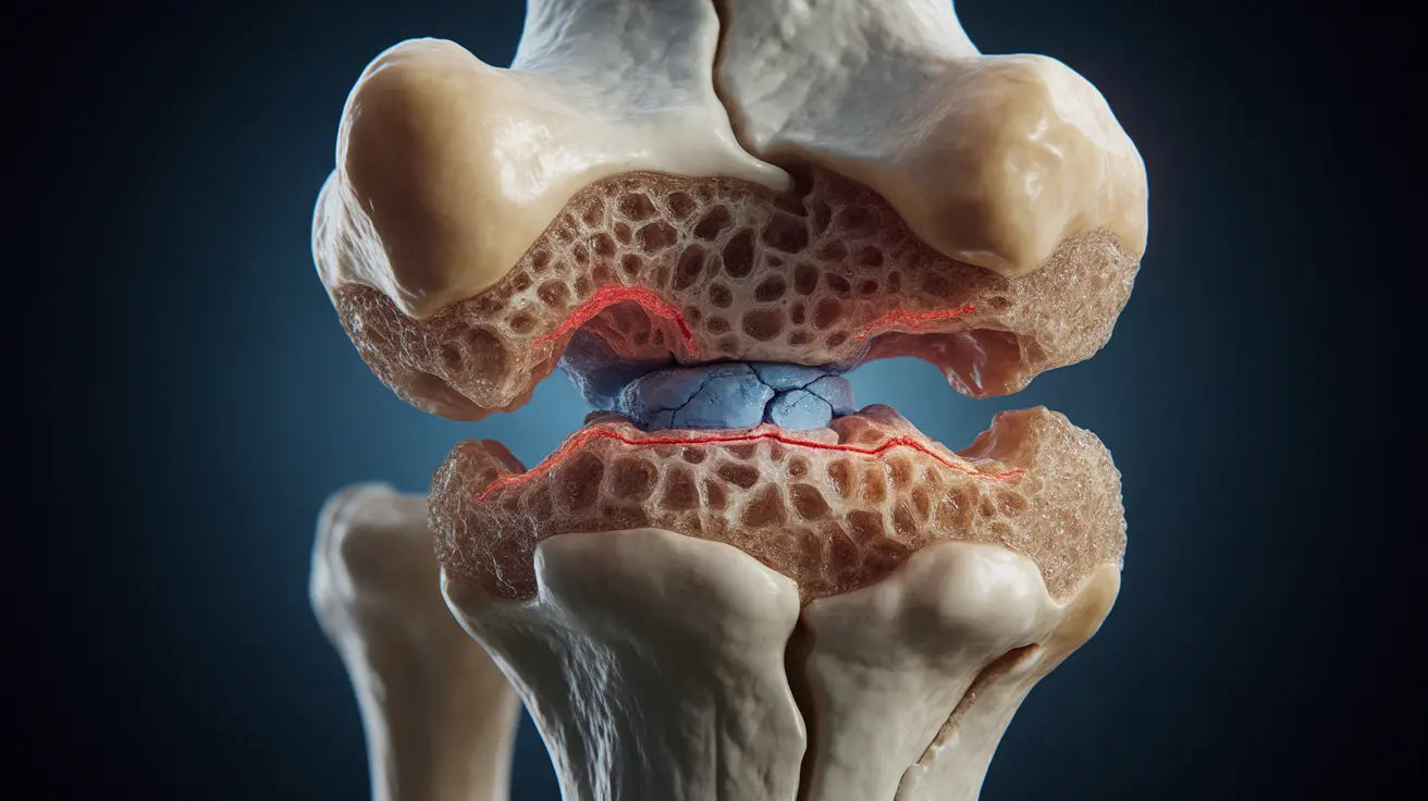
A subchondral fracture occurs in the layer of bone directly beneath joint cartilage, potentially causing significant pain and mobility issues. These fractures commonly affect weight-bearing joints like the knee and hip, particularly in individuals with underlying bone conditions or those who engage in high-impact activities. Understanding the nature of these fractures is crucial for proper diagnosis and treatment.
While sometimes confused with other joint conditions, subchondral fractures require specific medical attention and management strategies. This comprehensive guide explores the key aspects of subchondral fractures, from recognition to recovery.
Signs and Symptoms of Subchondral Fractures
Recognizing the symptoms of a subchondral fracture is essential for seeking timely medical attention. Common indicators include:
- Sudden, severe joint pain
- Pain that worsens with weight-bearing activities
- Localized swelling and tenderness
- Reduced range of motion
- Difficulty performing daily activities
The intensity of symptoms can vary significantly among individuals, often depending on the fracture's location and severity. Some people may experience gradual onset of pain, while others might notice sudden, acute discomfort.
Diagnostic Approaches
Diagnosing a subchondral fracture often requires multiple imaging techniques, as early signs may not be visible on standard X-rays. Healthcare providers typically use:
Advanced Imaging Methods
- MRI scans (most effective for early detection)
- CT scans for detailed bone structure analysis
- Bone scans in specific cases
- High-resolution X-rays for follow-up monitoring
Early diagnosis is crucial for preventing further joint damage and implementing appropriate treatment strategies. Healthcare providers will also conduct physical examinations and review medical history to ensure accurate diagnosis.
Treatment Strategies and Management
Treatment approaches for subchondral fractures vary based on severity, location, and individual patient factors. Common treatment options include:
Conservative Treatment
- Rest and activity modification
- Protected weight-bearing with assistive devices
- Physical therapy exercises
- Pain management medications
Surgical Interventions
Surgery may be necessary in cases where conservative treatment proves insufficient. Surgical options might include:
- Bone grafting
- Internal fixation
- Joint replacement in severe cases
Risk Factors and Prevention
Understanding risk factors is crucial for prevention and management of subchondral fractures. Key risk factors include:
- Osteoporosis and low bone density
- Advanced age
- Female gender, particularly post-menopausal women
- History of joint trauma
- High-impact athletic activities
Frequently Asked Questions
What are the common symptoms and signs of a subchondral fracture in weight-bearing joints like the knee or hip? The most common symptoms include acute joint pain, especially during weight-bearing activities, localized swelling, reduced mobility, and tenderness around the affected joint. Pain typically worsens with activity and improves with rest.
How is a subchondral fracture diagnosed, especially when early symptoms may not show on X-rays? Diagnosis typically involves multiple imaging techniques, with MRI being the gold standard for early detection. Healthcare providers may also use CT scans, bone scans, and detailed physical examinations to confirm the diagnosis and assess the extent of the injury.
What are the main treatment options for subchondral insufficiency fractures and when is surgery considered? Treatment options range from conservative approaches like protected weight-bearing and physical therapy to surgical interventions. Surgery is typically considered when conservative treatments fail to provide relief or in cases of severe fractures that threaten joint stability.
Can osteoporosis or low bone density increase the risk of developing a subchondral fracture? Yes, osteoporosis and low bone density significantly increase the risk of subchondral fractures. These conditions weaken bone structure, making it more susceptible to stress and injury, particularly in weight-bearing joints.
How can weight-bearing and physical activity be managed to help heal or prevent subchondral fractures? Management typically involves a gradual return to weight-bearing activities under medical supervision. This may include using assistive devices, following a structured physical therapy program, and avoiding high-impact activities during healing. Prevention focuses on maintaining bone health through appropriate exercise and nutrition.


