


Skin cancer is the most common form of cancer in the United States, with over 5 million cases diagnosed annually. Early detection can make the difference between a minor procedure and a life-threatening condition, which is why understanding the visual characteristics of different skin cancer types is crucial for everyone.
Learning to identify suspicious changes in moles, spots, and skin lesions empowers you to seek timely medical attention. While self-examination is valuable, it's important to remember that only a qualified dermatologist can provide a definitive diagnosis through proper examination and, if necessary, biopsy.
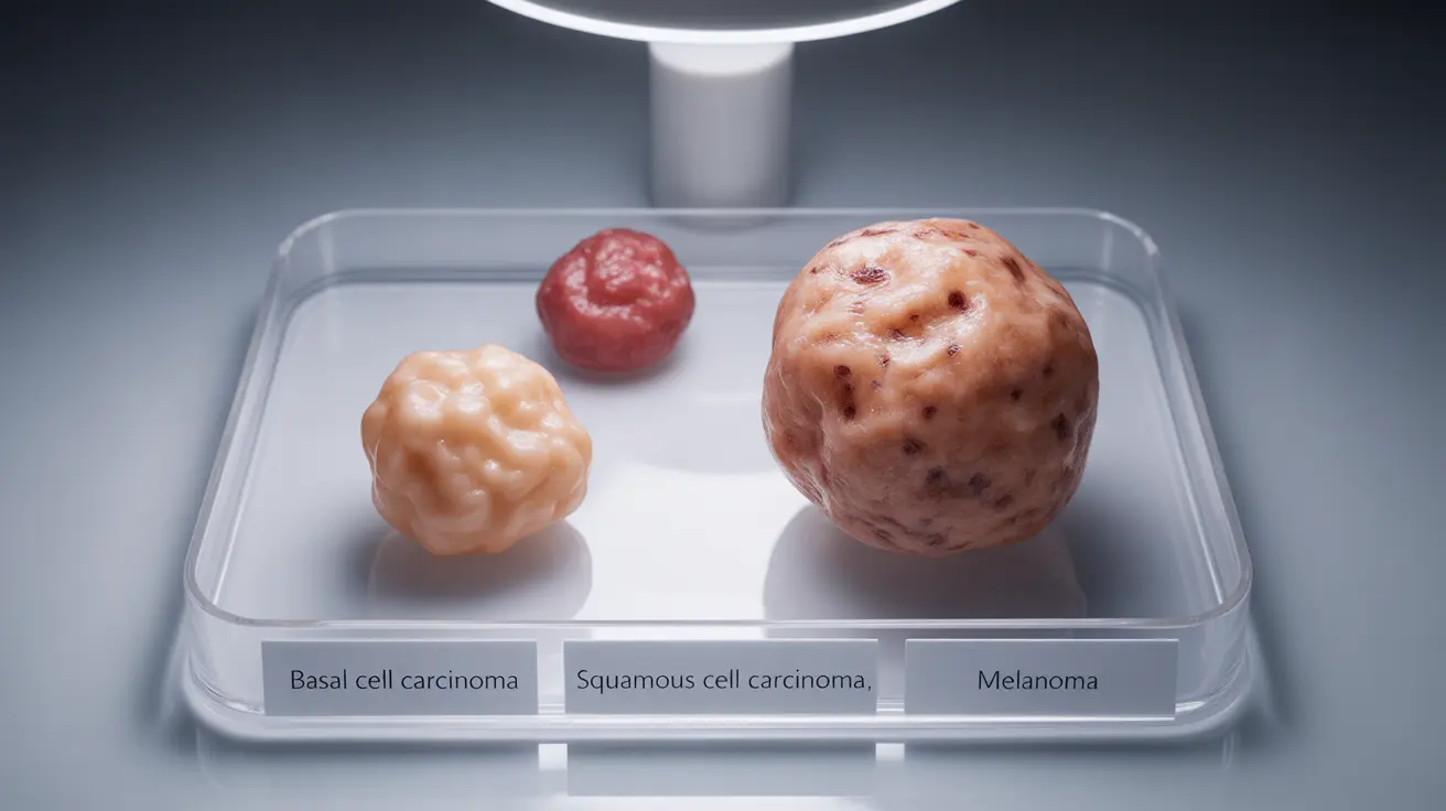
Understanding the Three Main Types of Skin Cancer
Skin cancer develops when skin cells undergo abnormal changes, typically due to DNA damage from ultraviolet radiation. The three primary categories each have distinct visual characteristics that can help with early identification.
Basal Cell Carcinoma: The Most Common Type
Basal cell carcinoma accounts for approximately 80% of all skin cancer diagnoses. This type typically develops in areas frequently exposed to sun, particularly the face, neck, and hands. Unlike other forms, basal cell carcinoma rarely spreads to other parts of the body, making it highly treatable when caught early.
Visually, basal cell carcinoma can appear as a pearly or waxy bump with a translucent quality. Some lesions present as flat, flesh-colored or brown scar-like areas, while others may look like open sores that don't heal properly or repeatedly bleed and scab over.
Squamous Cell Carcinoma: The Second Most Common
Squamous cell carcinoma represents about 20% of skin cancer cases and typically appears on sun-exposed areas including the face, ears, neck, lips, and backs of hands. This type has a higher potential to spread compared to basal cell carcinoma, particularly if left untreated.
These lesions often manifest as firm, red nodules or flat lesions with a scaly, crusted surface. They may resemble warts or open sores that don't heal within several weeks. Some people notice persistent tenderness or pain in the affected area.
Melanoma: The Most Serious Form
Although melanoma accounts for only about 1% of skin cancer cases, it causes the majority of skin cancer deaths due to its aggressive nature and tendency to spread quickly. Melanoma can develop anywhere on the body, including areas not typically exposed to sun.
Melanoma often develops from existing moles or appears as new, unusual-looking growths. The key to identification lies in recognizing changes in size, shape, color, or texture of existing moles, or the appearance of new spots that look different from other moles on your body.
The ABCDE Method for Melanoma Detection
Dermatologists recommend the ABCDE method as a systematic approach to evaluate suspicious moles and skin spots. This acronym helps identify the key warning signs that warrant professional evaluation.
A - Asymmetry
Normal moles are typically symmetrical, meaning one half mirrors the other. Melanomas often display asymmetry, where one half of the mole looks different from the other half when divided by an imaginary line.
B - Border Irregularity
Benign moles usually have smooth, even borders. Suspicious lesions may have irregular, scalloped, or poorly defined edges that appear ragged or blurred.
C - Color Variation
Healthy moles maintain consistent coloring throughout. Warning signs include multiple colors within a single mole, such as various shades of brown, black, red, white, or blue.
D - Diameter
While melanomas can be smaller, lesions larger than 6 millimeters (about the size of a pencil eraser) require closer attention. However, don't ignore smaller spots that exhibit other warning signs.
E - Evolution
Any mole that changes in size, shape, color, elevation, or develops new symptoms like bleeding, itching, or crusting should be evaluated by a healthcare professional promptly.
Recognizing Other Warning Signs
Beyond the ABCDE criteria, several additional symptoms can indicate skin cancer. Persistent sores that don't heal within four to six weeks often signal basal or squamous cell carcinoma. Recurring bleeding from a skin spot, especially without obvious injury, warrants medical attention.
Changes in sensation, such as itching, tenderness, or pain in a mole or skin lesion, can also indicate malignant transformation. Some people notice a spreading of pigment from a mole into surrounding skin, creating an irregular appearance.
New growths that appear different from other moles on your body, often called "ugly duckling" lesions, deserve professional evaluation even if they don't meet traditional criteria for concern.
When to See a Dermatologist
Schedule a dermatological examination if you notice any changes in existing moles or the appearance of new, unusual spots. Annual skin checks are recommended for individuals with risk factors including fair skin, history of sunburns, family history of skin cancer, or numerous moles.
Don't delay seeking medical attention for spots that bleed, itch, or change rapidly. Early-stage skin cancers are highly curable, with five-year survival rates exceeding 99% for localized melanomas and nearly 100% for basal and squamous cell carcinomas.
Frequently Asked Questions
What are the different types of skin cancer and how do they look different from each other?
The three main types of skin cancer have distinct appearances: Basal cell carcinoma typically appears as pearly, waxy bumps or flat, scar-like areas that may bleed easily. Squamous cell carcinoma presents as firm, red nodules or flat, scaly lesions that may resemble warts. Melanoma often looks like an irregular mole with asymmetrical shape, irregular borders, multiple colors, large diameter, or changes over time.
How can I tell the difference between a normal mole and melanoma using the ABCDE method?
The ABCDE method helps identify concerning features: A (Asymmetry) - one half doesn't match the other; B (Border) - irregular or poorly defined edges; C (Color) - multiple colors or uneven color distribution; D (Diameter) - larger than 6mm or growing; E (Evolution) - any changes in size, shape, color, elevation, or new symptoms like bleeding or itching. Normal moles are typically symmetrical, have even borders, consistent color, stable size, and remain unchanged over time.
What does basal cell carcinoma look like and where is it most likely to appear on my body?
Basal cell carcinoma commonly appears on sun-exposed areas like the face, neck, scalp, shoulders, and back. It typically looks like a pearly or waxy bump that may be translucent, or a flat, flesh-colored or brown scar-like area. Some appear as open sores that don't heal, bleed easily, or repeatedly scab and re-open. The lesions often have visible blood vessels and may have a central depression or ulceration.
Should I be worried about a spot on my skin that won't heal or keeps bleeding?
Yes, persistent sores that don't heal within 4-6 weeks or spots that bleed repeatedly without obvious injury should be evaluated by a healthcare professional promptly. These are common signs of basal cell or squamous cell carcinoma. Even minor bleeding from a mole or skin spot can indicate malignant changes and warrants medical attention, as early detection significantly improves treatment outcomes.
What are the warning signs that a skin lesion might be cancerous and when should I see a dermatologist?
Key warning signs include: changes in size, shape, color, or texture of existing moles; new growths that look different from other spots; persistent sores that don't heal; recurring bleeding; itching, tenderness, or pain; and asymmetrical or irregularly bordered lesions. See a dermatologist immediately if you notice any of these changes, or schedule annual skin checks if you have risk factors like fair skin, history of sunburns, family history of skin cancer, or numerous moles.


