


When doctors suspect Crohn's disease, they often recommend a biopsy as part of the diagnostic process. This procedure, while sometimes concerning to patients, provides crucial information that other tests simply cannot deliver. A Crohn's disease biopsy involves taking small tissue samples from the digestive tract to examine under a microscope, offering doctors a detailed view of inflammation patterns and cellular changes that are characteristic of this chronic inflammatory bowel condition.
Understanding why biopsies are necessary and what the process involves can help ease anxiety and ensure you're prepared for this important diagnostic step. While imaging tests and blood work provide valuable information, a biopsy remains one of the most definitive tools for confirming a Crohn's disease diagnosis and ruling out other similar conditions.
Why Biopsies Are Essential for Crohn's Disease Diagnosis
Imaging tests like CT scans and MRIs can reveal structural abnormalities and inflammation in the digestive tract, but they cannot provide the microscopic detail needed for a definitive Crohn's disease diagnosis. A biopsy allows doctors to examine the actual tissue architecture and identify specific cellular patterns that distinguish Crohn's disease from other inflammatory bowel conditions like ulcerative colitis or infectious colitis.
The microscopic examination reveals critical information about the depth and pattern of inflammation. Crohn's disease characteristically affects all layers of the intestinal wall, creating what doctors call "transmural inflammation." This deep, patchy inflammation pattern is a hallmark feature that can only be confirmed through tissue examination.
Additionally, biopsies help rule out other conditions that may mimic Crohn's disease symptoms, including certain infections, cancers, or other inflammatory conditions. This differential diagnosis is crucial for ensuring patients receive the most appropriate treatment plan.
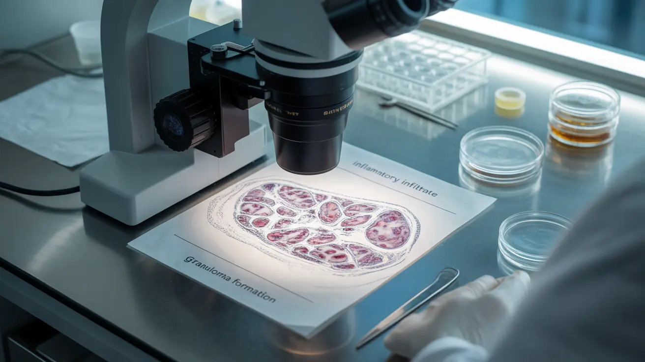
What Doctors Look for Under the Microscope
When examining Crohn's disease biopsy samples, pathologists search for several specific microscopic features that indicate the presence of this condition. The most significant finding is the presence of granulomas – small clusters of inflammatory cells that form in response to chronic irritation. However, granulomas are only found in about 25-50% of Crohn's disease cases, so their absence doesn't rule out the diagnosis.
Pathologists also examine the pattern and depth of inflammation throughout the tissue layers. In Crohn's disease, inflammation typically extends through all layers of the intestinal wall, from the innermost mucosa to the outermost serosa. This transmural inflammation creates characteristic changes in tissue architecture that experienced pathologists can readily identify.
Other microscopic features include increased numbers of inflammatory cells, particularly lymphocytes and plasma cells, distortion of normal gland structures, and evidence of chronic inflammation such as fibrosis or scarring. The pathologist also looks for signs of dysplasia or malignancy, which can occasionally develop in areas of chronic inflammation.
The Biopsy Procedure During Colonoscopy
Most Crohn's disease biopsies are obtained during a colonoscopy, a procedure that allows doctors to visualize the inside of the colon and terminal ileum directly. During the procedure, which is typically performed under sedation, the gastroenterologist uses a flexible tube with a camera to navigate through the digestive tract.
When suspicious areas are identified, the doctor uses small forceps passed through the colonoscope to take tiny tissue samples. These samples, usually measuring only 2-3 millimeters, are painless to obtain because the intestinal lining lacks pain receptors. Most patients don't feel the biopsy being taken, especially when under sedation.
The procedure typically involves taking multiple samples from different areas, including both inflamed and normal-appearing tissue. This helps pathologists compare affected areas with healthy tissue and provides a more comprehensive picture of the disease pattern. The entire biopsy process adds only a few minutes to the overall colonoscopy procedure.
Alternative Diagnostic Approaches
While biopsy remains the gold standard for Crohn's disease diagnosis, doctors sometimes use other approaches when colonoscopy isn't feasible or appropriate. Blood tests can detect inflammatory markers and antibodies associated with Crohn's disease, though these aren't specific enough for definitive diagnosis.
Advanced imaging techniques like magnetic resonance enterography (MRE) or CT enterography can reveal small bowel inflammation and complications that colonoscopy might miss. These tests are particularly useful for examining areas of the small intestine that are difficult to reach during colonoscopy.
In some cases, doctors may use capsule endoscopy, where patients swallow a small camera that takes pictures as it travels through the digestive tract. However, this method doesn't allow for tissue sampling, so it's typically used as a complementary diagnostic tool rather than a replacement for biopsy.
Understanding Granulomas and Other Inflammatory Signs
Granulomas are perhaps the most specific microscopic finding in Crohn's disease, though they're not present in all cases. These small, organized collections of inflammatory cells form when the immune system attempts to isolate and contain perceived threats. In Crohn's disease, granulomas typically don't have the central necrosis (tissue death) seen in infectious conditions like tuberculosis.
Other inflammatory signs visible under the microscope include crypt distortion, where the normal tubular structures in the intestinal lining become irregular or branched. Increased inflammatory cell infiltration, particularly in the deeper tissue layers, is another key feature that distinguishes Crohn's disease from other conditions.
The pathologist also evaluates the overall architecture of the tissue, looking for signs of chronic inflammation such as increased fibrous tissue, thickened blood vessel walls, and changes in the nerve networks within the intestinal wall. These features help determine disease severity and duration.
Frequently Asked Questions
Why do doctors need to take a biopsy to diagnose Crohn's disease instead of just using imaging tests?
While imaging tests like CT scans and MRIs can show inflammation and structural changes in the digestive tract, they cannot provide the microscopic detail necessary for a definitive Crohn's disease diagnosis. Biopsies allow doctors to examine the specific patterns of inflammation, identify characteristic cellular changes, and rule out other conditions that may appear similar on imaging studies. The microscopic examination reveals transmural inflammation and other features unique to Crohn's disease that imaging alone cannot detect.
What exactly do doctors look for in a Crohn's disease biopsy under the microscope?
Pathologists examine biopsy samples for several key features including granulomas (small clusters of inflammatory cells), transmural inflammation extending through all layers of the intestinal wall, increased inflammatory cell infiltration, crypt distortion, and architectural changes in the tissue structure. They also look for signs of chronic inflammation such as fibrosis and evaluate the overall pattern of disease involvement to distinguish Crohn's disease from other inflammatory bowel conditions.
How is a biopsy taken during a colonoscopy, and does it hurt?
During colonoscopy, biopsies are obtained using small forceps passed through the flexible scope. The doctor takes tiny tissue samples (2-3 millimeters) from suspicious or inflamed areas. The procedure is painless because the intestinal lining lacks pain receptors, and most patients are under sedation during the procedure. Taking biopsies adds only a few minutes to the overall colonoscopy and doesn't cause discomfort or complications in the vast majority of cases.
Can Crohn's disease be diagnosed without a biopsy?
While biopsy remains the gold standard for Crohn's disease diagnosis, doctors sometimes make clinical diagnoses based on a combination of symptoms, blood tests, imaging studies, and endoscopic findings. However, this approach is less definitive and may lead to diagnostic uncertainty. In cases where colonoscopy isn't possible, doctors may use alternative methods like capsule endoscopy or advanced imaging, but tissue confirmation through biopsy provides the most accurate diagnosis and helps guide appropriate treatment decisions.
What is the difference between granulomas found in a biopsy and other signs of Crohn's disease inflammation?
Granulomas are specific, organized clusters of inflammatory cells that form in response to chronic irritation and are considered the most characteristic microscopic finding in Crohn's disease, though they're only present in 25-50% of cases. Other inflammatory signs include diffuse inflammatory cell infiltration throughout tissue layers, crypt distortion, architectural changes, and transmural inflammation patterns. While granulomas are highly specific for Crohn's disease when present, their absence doesn't rule out the diagnosis, and doctors rely on the overall pattern of inflammatory changes to make the diagnosis.


