


When diagnosing and monitoring uterine cancer, medical imaging plays a crucial role in determining the extent of the disease and planning appropriate treatment. CT scans with contrast are one of several important imaging tools that healthcare providers may use, particularly when evaluating if cancer has spread beyond the uterus.
This comprehensive guide explores how contrast-enhanced CT scans work in uterine cancer cases, their specific uses, and what patients can expect during the procedure. We'll also discuss how this imaging method compares to other diagnostic tools.
The Role of CT Scans in Uterine Cancer Management
CT scans with contrast are primarily used in uterine cancer cases to assess whether the cancer has spread to other parts of the body, rather than for initial diagnosis. The contrast material helps create clearer images of organs, blood vessels, and potential tumor sites.
How Contrast Enhancement Works
During a contrast-enhanced CT scan, a special dye is administered either orally or intravenously. This dye helps highlight specific areas of the body, making it easier for radiologists to identify potential cancer spread, lymph node involvement, or other abnormalities that might not be visible on a standard CT scan.
Applications and Limitations
While CT scans with contrast are valuable tools, they have specific applications and limitations in uterine cancer cases:
- Detecting cancer spread to distant organs
- Evaluating lymph node involvement
- Monitoring treatment response
- Planning surgical procedures
However, other imaging methods, such as transvaginal ultrasound and MRI, are typically preferred for initial diagnosis and local staging of uterine cancer.
The CT Scan Procedure
Understanding what happens during a CT scan with contrast can help patients feel more prepared:
Before the Scan
Patients may need to:
- Fast for several hours
- Remove metal objects and jewelry
- Inform their doctor about any allergies or kidney problems
- Sign consent forms for contrast administration
During the Scan
The actual scanning process typically takes 15-30 minutes. Patients lie still on a table that moves through a donut-shaped scanner while the contrast material provides enhanced imaging of internal structures.
CT Scans vs. Other Imaging Methods
Different imaging techniques serve various purposes in uterine cancer evaluation:
- CT scans: Best for detecting distant spread
- MRI: Superior for evaluating local disease extent
- Ultrasound: Typically used for initial evaluation
- PET scans: Sometimes combined with CT for comprehensive staging
Frequently Asked Questions
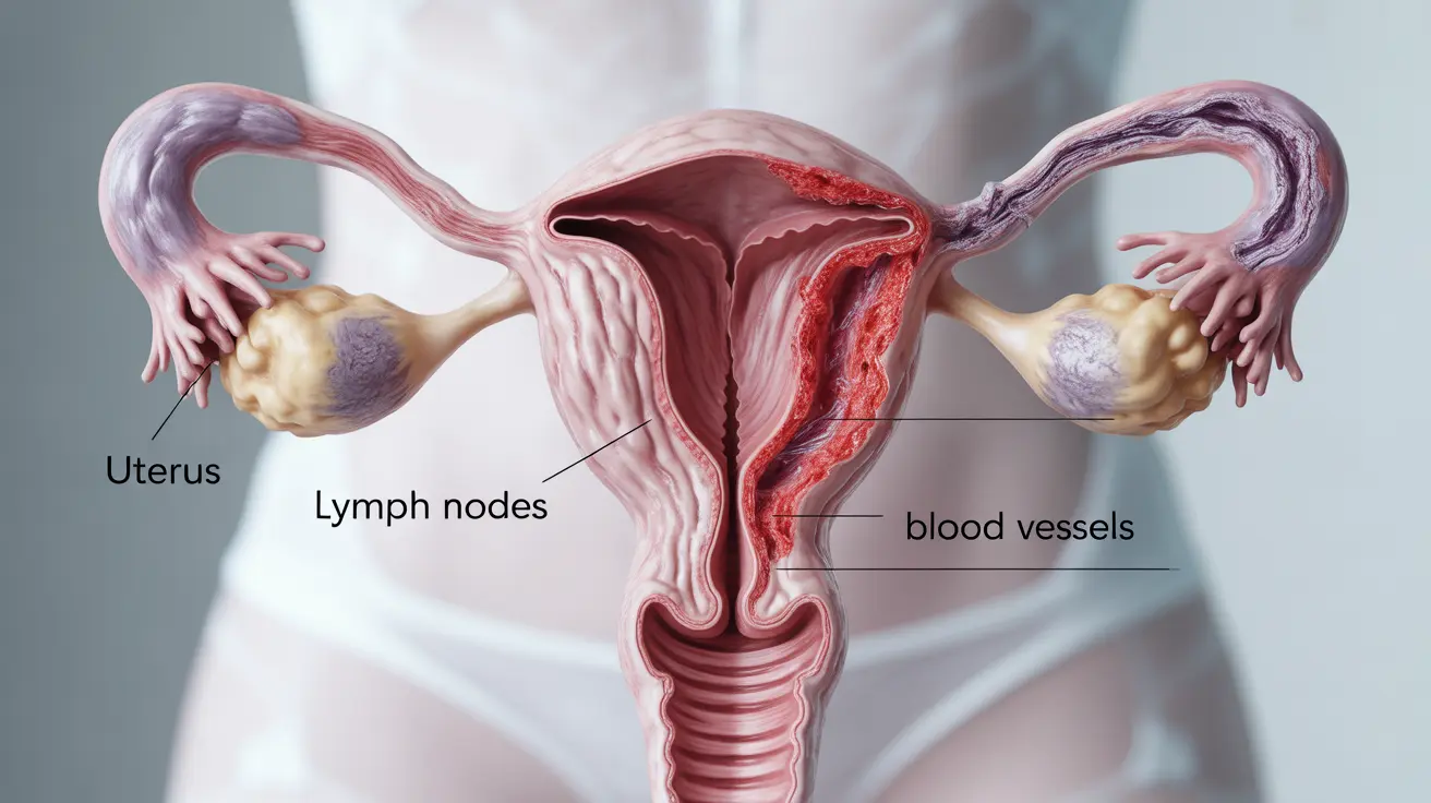
What does a CT scan with contrast show in uterine cancer patients? A CT scan with contrast can reveal whether cancer has spread beyond the uterus, showing potential involvement in lymph nodes, blood vessels, and distant organs. The contrast material helps create clearer images of these structures.
How accurate is a contrast-enhanced CT scan for detecting spread of uterine cancer? Contrast-enhanced CT scans are highly accurate for detecting cancer spread to distant organs and enlarged lymph nodes, with accuracy rates typically around 85-95% for detecting metastatic disease.
Why is a CT scan not usually used to diagnose uterine cancer initially? CT scans aren't typically used for initial diagnosis because other methods like transvaginal ultrasound and MRI provide better visualization of the uterine tissue itself. These methods are more effective for evaluating the local extent of the disease.
What should I expect during a CT scan with contrast for uterine cancer evaluation? During the procedure, you'll lie on a table that moves through a scanner. You'll receive contrast material either through an IV or orally. The scan typically takes 15-30 minutes, and you'll need to lie still throughout the procedure.
How does a CT scan compare to MRI when staging uterine cancer? While CT scans excel at detecting cancer spread to distant organs, MRI provides superior soft tissue contrast and is better at showing the depth of tumor invasion in the uterus. MRI is generally preferred for local staging, while CT is better for evaluating distant spread.


