


A chest X-ray plays a crucial role in diagnosing and screening for tuberculosis (TB), providing healthcare providers with valuable insights into potential lung infections. Understanding what this important diagnostic tool can reveal and what to expect during the procedure can help patients feel more prepared and informed about their TB testing journey.
This comprehensive guide explores how chest X-rays contribute to TB diagnosis, their effectiveness, limitations, and what you should know before getting one.
How Chest X-Rays Help Detect Tuberculosis
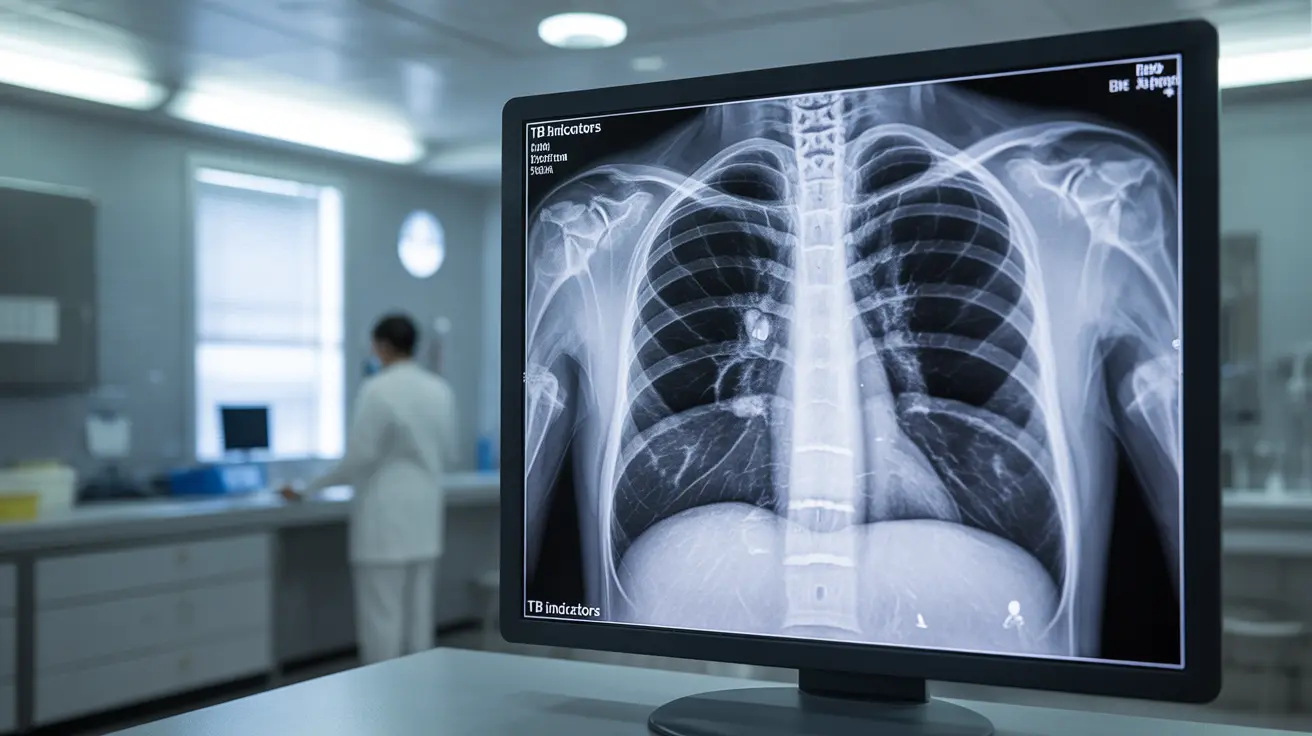
Chest X-rays create detailed images of your lungs, heart, and surrounding structures, allowing doctors to identify characteristic signs of TB infection. During interpretation, medical professionals look for specific patterns and abnormalities that might indicate the presence of tuberculosis.
Key Features Doctors Look For
When examining a chest X-ray for TB, doctors typically search for:
- Infiltrates or shadows in the upper lobes
- Cavity formations
- Enlarged lymph nodes
- Pleural effusions (fluid around the lungs)
- Miliary patterns indicating disseminated TB
- Calcified nodules from previous infections
The Role of Chest X-Rays in TB Diagnosis
While chest X-rays are valuable screening tools, they form just one part of a comprehensive TB diagnosis approach. They're particularly useful for:
- Initial screening in high-risk populations
- Monitoring treatment progress
- Identifying potential complications
- Following up on positive skin tests
- Screening before starting certain medications
Limitations of X-Ray Testing
Despite their utility, chest X-rays have certain limitations in TB diagnosis. They cannot definitively confirm TB on their own, as other conditions may present similar radiographic patterns. Additionally, early-stage TB might not show clear signs on an X-ray, necessitating additional testing methods.
Preparing for Your TB Chest X-Ray
Getting ready for a chest X-ray is relatively straightforward. You'll need to:
- Remove any jewelry or metal objects from your chest area
- Wear comfortable clothing that's easy to remove
- Inform your healthcare provider about any recent illnesses
- Mention if you're pregnant or might be pregnant
- Follow any specific instructions provided by your healthcare team
Safety and Radiation Exposure
Chest X-rays are considered safe diagnostic procedures with minimal radiation exposure. The benefits of detecting potential TB infection generally outweigh the very small risks associated with radiation exposure from a single chest X-ray.
Frequently Asked Questions
How does a chest X-ray help diagnose active tuberculosis and what signs do doctors look for? Doctors examine chest X-rays for specific patterns including upper lobe infiltrates, cavities, and lymph node enlargement. These signs can indicate active TB infection, though additional tests are usually needed for confirmation.
Can a chest X-ray detect latent tuberculosis infection or only active TB? Chest X-rays primarily detect active TB disease. Latent TB typically doesn't show visible changes on X-rays, which is why other tests like skin or blood tests are necessary for detecting latent infection.
What are the limitations of using chest X-rays alone in diagnosing tuberculosis? X-rays alone cannot definitively diagnose TB as other conditions may show similar patterns. They also might miss early infections or show unclear results in some cases, requiring additional diagnostic methods for confirmation.
When is a chest X-ray recommended as part of tuberculosis testing or screening? Chest X-rays are recommended after positive skin tests, for initial screening in high-risk populations, during regular monitoring of TB treatment, and when TB symptoms are present.
Is a chest X-ray safe and how should I prepare for a TB-related chest X-ray exam? Yes, chest X-rays are safe with minimal radiation exposure. Preparation involves removing metal objects and jewelry from the chest area, wearing comfortable clothing, and informing healthcare providers about potential pregnancy or recent illnesses.


